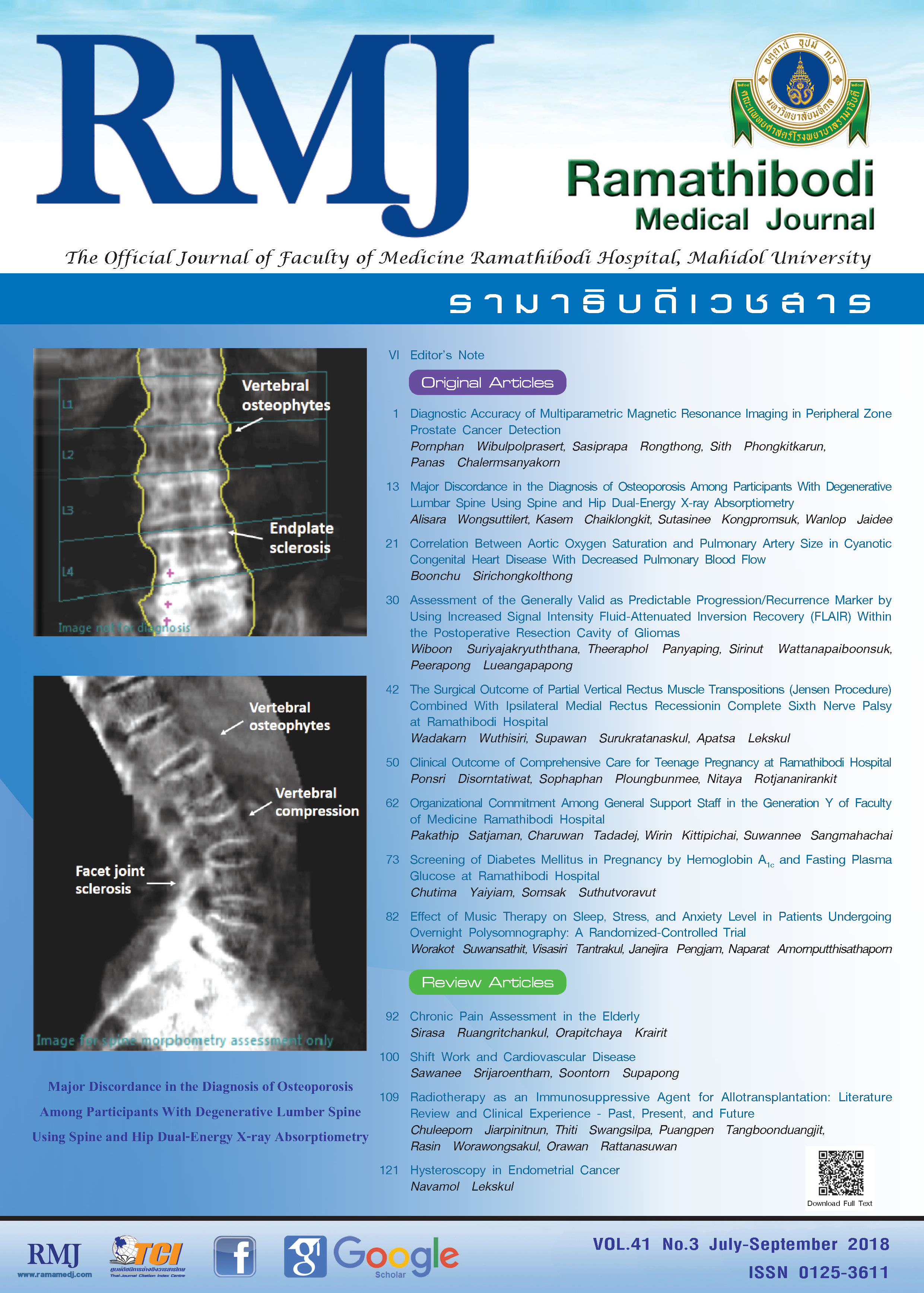
Major Discordance in the Diagnosis of Osteoporosis Among Participants With Degenerative Lumbar Spine Using Spine and Hip Dual-Energy X-ray Absorptiometry
DOI:
https://doi.org/10.14456/rmj.2018.33Keywords:
Bone mineral density, DXA, Lumbar spine, Degenerative spine diseaseAbstract
Background: Major T-score discordance in the diagnosis of osteoporosis at spine and the hip may induce misinterpretation in people with degenerative lumbar spine.
Objective: To investigate major T-score discordance in the diagnosis of osteoporosis using bone mineral density (BMD) at the lumbar spine and the hip among people with degenerative lumbar spine.
Methods: Demographic data, anthropometric measurements, and dual-energy X-ray absorptiometry (DXA) scans at the lumbar spine and the hip were retrieved from a database of Burapha University Hospital. The interrater reliabilities of image interpretation were performed. The correlation between degenerative lumbar spine lesions with T-scores at the lumbar spine and the hip was analyzed. Major discordance was defined when one site is osteoporotic and the other site is osteopenia or normal.
Results: Major T-score discordance of the lumbar spine and the hip was 28.6% and the hip BMD detected in participants with osteoporosis was less than the lumbar spine BMD (15.7% vs 36.5%). T-score at lumbar spine showed a false negative rate of 25.0%. Degenerative lumbar spine lesions showed a poor correlation with T-score at the lumbar spine (R2 = 0.21, P < 0.01) and the hip (R2 at femoral neck = 0.24, P < 0.01; R2 at total hip = 0.32, P < 0.01).
Conclusions: Twenty-six percent of participants with degenerative lumbar spine were not evaluated L1-L4 BMD. While the hip seems to be the best measurement site, 28.6% of T-score discordance between lumbar spine and hip was still observed. T-score at lumbar spine showed a false negative rate of 25.0% and associated with older age. This study suggested that it would not be suitable to use the hip BMD alone to diagnose osteoporosis in older adults with degenerative spine disease.
References
Jain RK, Vokes T. Dual-energy X-ray absorptiometry. J Clin Densitom. 2017;20(3):291-303. doi:10.1016/j.jocd.2017.06.014.
ISCD. Official Positions-Adult, 2015. International Society for Clinical Densitometry ISCD). https://www.iscd.org/official-positions/2015-iscd-official-positions-adult/. Accessed May 27, 2018.
Rand T, Seidl G, Kainberger F, et al. Impact of spinal degenerative changes on the evaluation of bone mineral density with dual energy X-ray absorptiometry (DXA). Calcif Tissue Int. 1997;60(5):430-433.
Muraki S, Yamamoto S, Ishibashi H, et al. Impact of degenerative spinal diseases on bone mineral density of the lumbar spine in elderly women. Osteoporos Int. 2004;15(9):724-728. doi:10.1007/s00198-004-1600-y.
Schneider DL, Bettencourt R, Barrett-Connor E. Clinical utility of spine bone density in elderly women. J Clin Densitom. 2006;9(3):255-260. doi:10.1016/j.jocd.2006.04.116.
Tenne M, McGuigan F, Besjakov J, Gerdhem P, Åkesson K. Degenerative changes at the lumbar spine--implications for bone mineral density measurement in elderly women. Osteoporos Int. 2013;24(4):1419-28. doi:10.1007/s00198-012-2048-0.
Moayyeri A, Soltani A, Bahrami H, Sadatsafavi M, Jalili M, Larijani B. Preferred skeletal site for osteoporosis screening in high-risk populations. Public Health. 2006;120(9):863-871. doi:10.1016/j.puhe.2006.05.013.
Atalay A, Kozakcioglu M, Cubuk R, Tasali N, Guney S. Degeneration of the lumbar spine and dual-energy X-ray absorptiometry measurements in patients without osteoporosis. Clin Imaging. 2009;33(5):374-378. doi:10.1016/j.clinimag.2008.12.005.
Krege JH, Miller PD, Lenchik L, Misurski DA, Chen P. New or worsening lumbar spine vertebral fractures increase lumbar spine bone mineral density and falsely suggest improved skeletal status. J Clin Densitom. 2006;9(2):144-149. doi:10.1016/j.jocd.2006.02.001.
Bonnick SL, Lewis LA. Bone Densitometry for Technologists. 3rd ed. New York, NY: Springer; 2013.
El Maghraoui A, Mouinga Abayi DA, Rkain H, Mounach A. Discordance in diagnosis of osteoporosis using spine and hip bone densitometry. J Clin Densitom. 2007;10(2):153-156. doi:10.1016/j.jocd.2006.12.003.
World Health Organization. WHO Expert Committee on Physical Status: the Use and Interpretation of Anthropometry. Geneva, Switzerland: World Health Organization; 1995. https://apps.who.int/iris/bitstream/handle/10665/37003/WHO_TRS_854.pdf?sequence=1&isAllowed=y. Accessed May 27, 2018.
Genant HK, Wu CY, van Kuijk C, Nevitt MC. Vertebral fracture assessment using a semiquantitative technique. J Bone Miner Res. 1993;8(9):1137-1148. doi:10.1002/jbmr.5650080915.
Landis JR, Koch GG. The measurement of observer agreement for categorical data. Biometrics. 1977;33(1):159-174.
Moayyeri A, Soltani A, Tabari NK, Sadatsafavi M, Hossein-Neghad A, Larijani B. Discordance in diagnosis of osteoporosis using spine and hip bone densitometry. BMC Endocr Disord. 2005;5(1):3. doi:10.1186/1472-6823-5-3.
Mounach A, Abayi DA, Ghazi M, et al. Discordance between hip and spine bone mineral density measurement using DXA: prevalence and risk factors. Semin Arthritis Rheum. 2009;38(6):467-471. doi:10.1016/j.semarthrit.2008.04.001.
Singh M, Magon N, Singh T. Major and minor discordance in the diagnosis of postmenopausal osteoporosis among Indian women using hip and spine dual-energy X-ray absorptiometry. J Midlife Health. 2012;3(2):76-80. doi:10.4103/0976-7800.104457.
Ikegami S, Kamimura M, Uchiyama S, et al. Bone mineral density measurement at both spine and hip for diagnosing osteoporosis in Japanese patients. J Clin Densitom. 2009;12(3):337-344. doi:10.1016/j.jocd.2009.03.099.
Faulkner KG, von Stetten E, Miller P. Discordance in patient classification using T-scores. J Clin Densitom. 1999;2(3):343-350.
Woodson G. Dual X-ray absorptiometry T-score concordance and discordance between the hip and spine measurement sites. J Clin Densitom. 2000;3(4):319-324.
O'Gradaigh D, Debiram I, Love S, Richards HK, Compston JE. A prospective study of discordance in diagnosis of osteoporosis using spine and proximal femur bone densitometry. Osteoporos Int. 2003;14(1):13-18. doi:10.1007/s00198-002-1311-1.
Carey JJ, Delaney MF, Love TE, et al. DXA-generated Z-scores and T-scores may differ substantially and significantly in young adults. J Clin Densitom. 2007;10(4):351-358. doi:10.1016/j.jocd.2007.06.001.
Callréus M, McGuigan F, Akesson K. Country-specific young adult dual-energy X-ray absorptiometry reference data are warranted for T-score calculations in women: data from the peak-25 cohort. J Clin Densitom. 2014;17(1):129-135. doi:10.1016/j.jocd.2013.03.008.
Pye SR, Reid DM, Adams JE, Silman AJ, O'Neill TW. Radiographic features of lumbar disc degeneration and bone mineral density in men and women. Ann Rheum Dis. 2006;65(2):234-238. doi:10.1136/ard.2005.038224.


