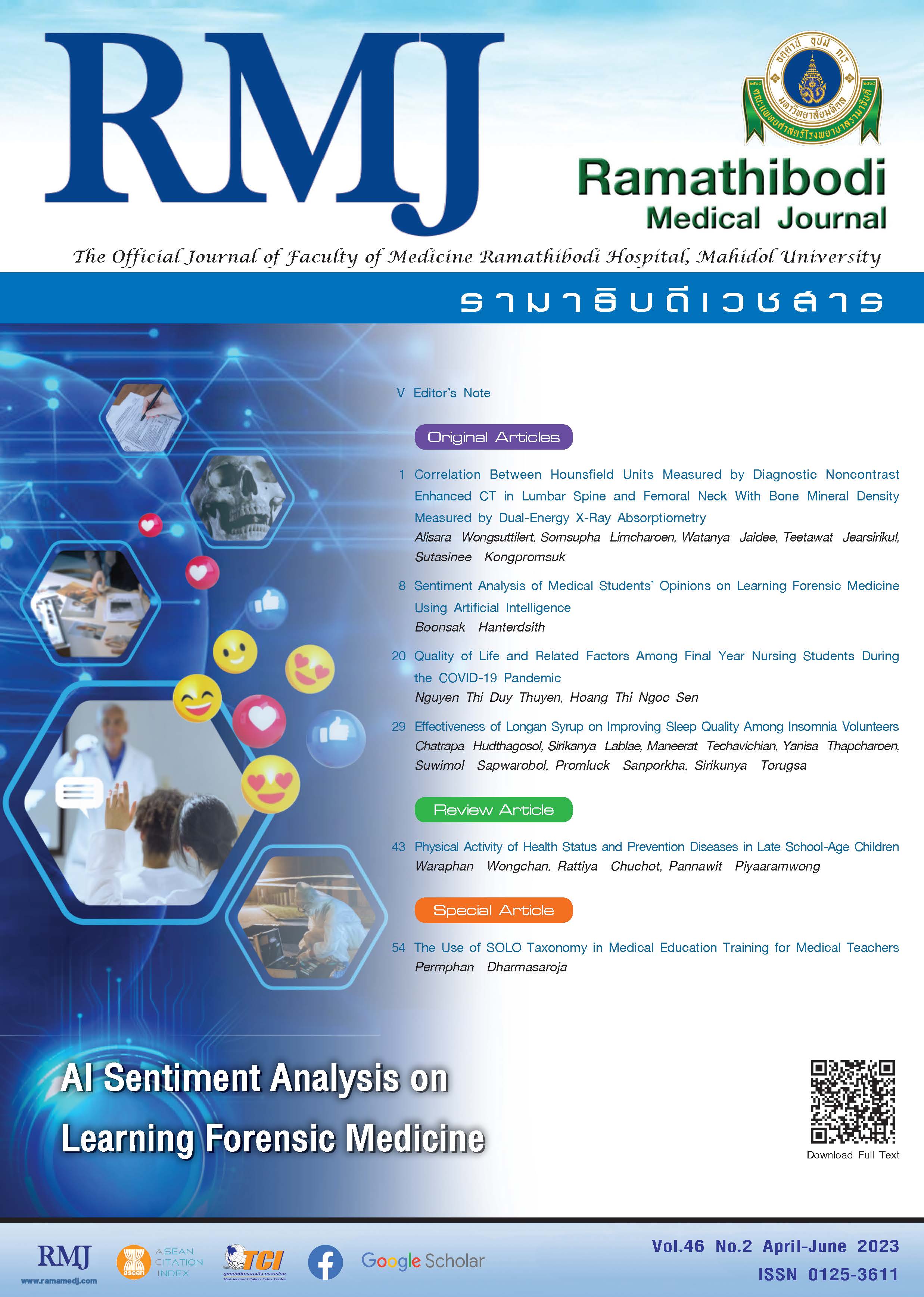
Correlation Between Hounsfield Units Measured by Diagnostic Noncontrast Enhanced CT in Lumbar Spine and Femoral Neck With Bone Mineral Density Measured by Dual-Energy X-Ray Absorptiometry
DOI:
https://doi.org/10.33165/rmj.2023.46.2.262589Keywords:
DXA, Bone density, CT scan, OsteoporosisAbstract
Background: Computed tomography (CT) is widely available and measurement of Hounsfield unit (HU) is easy and reproducible. Can this measurement predict osteoporosis?
Objective: To evaluate correlation between HU of the lumbar spine and femoral neck and bone mineral density T-score (BMD T-score), and to assess the cutoff HU values at the lumbar spine and femoral neck to predict osteoporosis.
Methods: This retrospective study reviewed 237 patients who underwent both dual-energy x-ray absorptiometry (DXA) and CT within an interval of 1 year from January 2014 to August 2022. HU at the L1-L4 lumbar spine and femoral neck, as well as BMD T-score for the same regions were determined. Correlation of BMD T-score and HU was evaluated using Pearson correlation. Receiver operating characteristic (ROC) curves were generated to determine the cutoff HU values for predicting osteoporosis and its sensitivity, specificity, positive, and negative predictive values.
Results: Among 205 patients included, 64 (31.2%) had osteoporosis. At the lumbar spine, the average HU was moderately correlated with the BMD T-score (r = 0.639; 95% CI, 0.550 - 0.714), whereas at the femoral neck, the average HU had a good correlation with BMD T-score (r = 0.756; 95% CI, 0.691 - 0.809). Cutoff values for predicting osteoporosis were 155 HU at the lumbar spine and 130 HU at the femoral neck.
Conclusions: HU and BMD T-score were more strongly correlated at the femoral neck than at the lumbar spine. A cutoff HU value of 130 at the femoral neck was used for predicting osteoporosis in Thai people.
References
Sözen T, Özışık L, Başaran NÇ. An overview and management of osteoporosis. Eur J Rheumatol. 2017;4(1):46-56. doi:10.5152/eurjrheum.2016.048
Camacho PM, Petak SM, Binkley N, et al. American Association of Clinical Endocrinologists/American College of Endocrinology clinical practice guidelines for the diagnosis and treatment of postmenopausal osteoporosis-2020 update. Endocr Pract. 2020;26(Suppl 1):1-46. doi:10.4158/GL-2020-0524SUPPL
Damilakis J, Adams JE, Guglielmi G, Link TM. Radiation exposure in x-ray-based imaging techniques used in osteoporosis. Eur Radiol. 2010;20(11):2707-2714. doi:10.1007/s00330-010-1845-0
Alawi M, Begum A, Harraz M, et al. Dual-energy x-ray absorptiometry (DEXA) scan versus computed tomography for bone density assessment. Cureus. 2021;13(2):e13261. doi:10.7759/cureus.13261
Islamian JP, Garoosi I, Fard KA, Abdollahi MR. Comparison between the MDCT and the DXA scanners in the evaluation of BMD in the lumbar spine densitometry. Egypt J Radiol Nucl Med. 2016;47(3):961-967. doi:10.1016/j.ejrnm.2016.04.005
Pickhardt PJ, Lee LJ, del Rio AM, et al. Simultaneous screening for osteoporosis at CT colonography: bone mineral density assessment using MDCT attenuation techniques compared with the DXA reference standard. J Bone Miner Res. 2011;26(9):2194-2203. doi:10.1002/jbmr.428
Link TM, Koppers BB, Licht T, Bauer J, Lu Y, Rummeny EJ. In vitro and in vivo spiral CT to determine bone mineral density: initial experience in patients at risk for osteoporosis. Radiology. 2004;231(3):805-811. doi:10.1148/radiol.2313030325
Kim KJ, Kim DH, Lee JI, Choi BK, Han IH, Nam KH. Hounsfield units on lumbar computed tomography for predicting regional bone mineral density. Open Med (Wars). 2019;14:545-551. doi:10.1515/med-2019-0061
Lee S, Chung CK, Oh SH, Park SB. Correlation between bone mineral density measured by dual-energy x-ray absorptiometry and Hounsfield units measured by diagnostic CT in lumbar spine. J Korean Neurosurg Soc. 2013;54(5):384-389. doi:10.3340/jkns.2013.54.5.384
Schreiber JJ, Anderson PA, Rosas HG, Buchholz AL, Au AG. Hounsfield units for assessing bone mineral density and strength: a tool for osteoporosis management. J Bone Joint Surg Am. 2011;93(11):1057-1063. doi:10.2106/JBJS.J.00160
Park H, Yang H, Heo J, et al. Bone mineral density screening interval and transition to osteoporosis in Asian women. Endocrinol Metab (Seoul). 2022;37(3):506-512. doi:10.3803/EnM.2022.1429
Hajian-Tilaki K. Sample size estimation in diagnostic test studies of biomedical informatics. J Biomed Inform. 2014;48:193-204. doi:10.1016/j.jbi.2014.02.013
Downloads
Published
How to Cite
Issue
Section
License
Copyright (c) 2023 Ramathibodi Medical Journal

This work is licensed under a Creative Commons Attribution-NonCommercial-NoDerivatives 4.0 International License.


