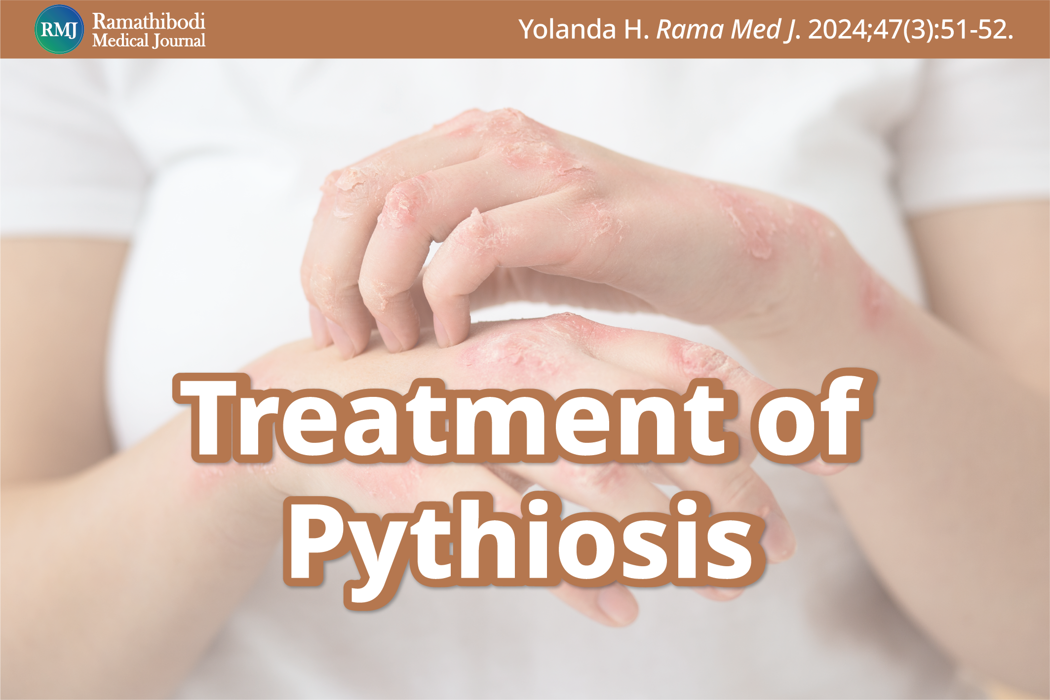
Treatment of Pythiosis
DOI:
https://doi.org/10.33165/rmj.2024.47.3.269238Abstract
Dear Editor: Due to its rising global incidence, I have dedicated years to studying pythiosis, a rare yet deadly infectious disease caused by Pythium insidiosum.1 Pythiosis typically presents with the infection of the artery, eye, gastrointestinal tract, and skin, and if left untreated, it can lead to organ loss or even death. In recent decades, a deeper understanding of P. insidiosum has been seen across molecular, serological, and histological studies, enhancing clinical awareness and diagnostic advancements.1,2 Traditionally mistaken for a fungus because of its microscopic appearance, P. insidiosum was treated with antifungal medications like terbinafine and amphotericin B.3,4 Molecular research clarifies that P. insidiosum is an oomycete, more closely related to algae, with significant biological differences from fungi, impacting drug efficacy.5 Since the 1980s, P. insidiosum antigen immunotherapy (PIAI) has emerged as a treatment alternative, with ongoing enhancements to increase efficacy.6 Surgery often serves as a last resort to limit the disease progression or to save lives.7
Current treatment strategies for pythiosis include antimicrobial drugs, PIAI, and surgery. Antimicrobial susceptibility testing reveals a higher sensitivity of P. insidiosum to antibacterials like macrolides, oxazolidinones, and tetracyclines, reducing the reliance on surgery.8 Though less effective, antifungals, sometimes in combination with antibacterials, can treat pythiosis.9,10 PIAI, prepared by crude antigen extract of P. insidiosum, is beneficial for humans and animals with pythiosis, potentially reducing surgery needs and increasing survival rates. However, its efficacy varies across different disease manifestations.6 Surgical intervention, typically reserved for unresponsive cases, ranges from organ-preserving procedures to more radical approaches like amputation, depending on disease progression.2,3,7 Additional treatments, including dimethyl sulfoxide, potassium iodide, steroids, ethanol, and mefenoxam, applied singly or combined, have shown promise in treating specific pythiosis forms.11,12
Treatment is the most challenging aspect of pythiosis, but there is hope. The morbidity and mortality of affected patients remain high. However, with continued attention and basic/clinical research by the medical community, we can gain insight into the disease and find a better way of pythiosis control, potentially improving their clinical outcomes.
References
Yolanda H, Krajaejun T. Global distribution and clinical features of pythiosis in humans and animals. J Fungi (Basel). 2022;8(2):182. doi:10.3390/jof8020182
Chitasombat MN, Jongkhajornpong P, Lekhanont K, Krajaejun T. Recent update in diagnosis and treatment of human pythiosis. PeerJ. 2020;8:e8555. doi:10.7717/peerj.8555
Krajaejun T, Sathapatayavongs B, Pracharktam R, et al. Clinical and epidemiological analyses of human pythiosis in Thailand. Clin Infect Dis. 2006;43(5):569-576. doi:10.1086/506353
Dória RG, Carvalho MB, Freitas SH, et al. Evaluation of intravenous regional perfusion with amphotericin B and dimethylsulfoxide to treat horses for pythiosis of a limb. BMC Vet Res. 2015;11:152. doi:10.1186/s12917-015-0472-z
Lerksuthirat T, Sangcakul A, Lohnoo T, Yingyong W, Rujirawat T, Krajaejun T. Evolution of the sterol biosynthetic pathway of Pythium insidiosum and related oomycetes contributes to antifungal drug resistance. Antimicrob Agents Chemother. 2017;61(4):10.1128/aac.02352-16. doi:10.1128/AAC.02352-16
Yolanda H, Krajaejun T. History and perspective of immunotherapy for pythiosis. Vaccines (Basel). 2021;9(10):1080. doi:10.3390/vaccines9101080
Permpalung N, Worasilchai N, Plongla R, et al. Treatment outcomes of surgery, antifungal therapy and immunotherapy in ocular and vascular human pythiosis: a retrospective study of 18 patients. J Antimicrob Chemother. 2015;70(6):1885-1892. doi:10.1093/jac/dkv008
Yolanda H, Krajaejun T. Review of methods and antimicrobial agents for susceptibility testing against Pythium insidiosum. Heliyon. 2020;6(4):e03737. doi:10.1016/j.heliyon.2020.e03737
Luangnara A, Chuamanochan M, Chiewchanvit S, Pattamapaspong N, Salee P, Chaiwarith R. Pythiosis presenting with chronic swelling and painful subcutaneous lesion at right deltoid. IDCases. 2023;33:e01873. doi:10.1016/j.idcr.2023.e01873
Torvorapanit P, Chuleerarux N, Plongla R, et al. Clinical outcomes of radical surgery and antimicrobial agents in vascular pythiosis: a multicenter prospective study. J Fungi (Basel). 2021;7(2):114. doi:10.3390/jof7020114
Yolanda H, Lohnoo T, Rujirawat T, et al. Selection of an appropriate in vitro susceptibility test for assessing anti-pythium insidiosum activity of potassium iodide, triamcinolone acetonide, dimethyl sulfoxide, and ethanol. J Fungi (Basel). 2022;8(11):1116. doi:10.3390/jof8111116
Billings P, Walton S, Shmalberg J, Santoro D. The use of mefenoxam to treat cutaneous and gastrointestinal pythiosis in dogs: a retrospective study. Microorganisms. 2023;11(7):1726. doi:10.3390/microorganisms11071726

Downloads
Published
How to Cite
Issue
Section
License
Copyright (c) 2024 by the Authors. Licensee Ramathibodi Medical Journal.

This work is licensed under a Creative Commons Attribution-NonCommercial-NoDerivatives 4.0 International License.


