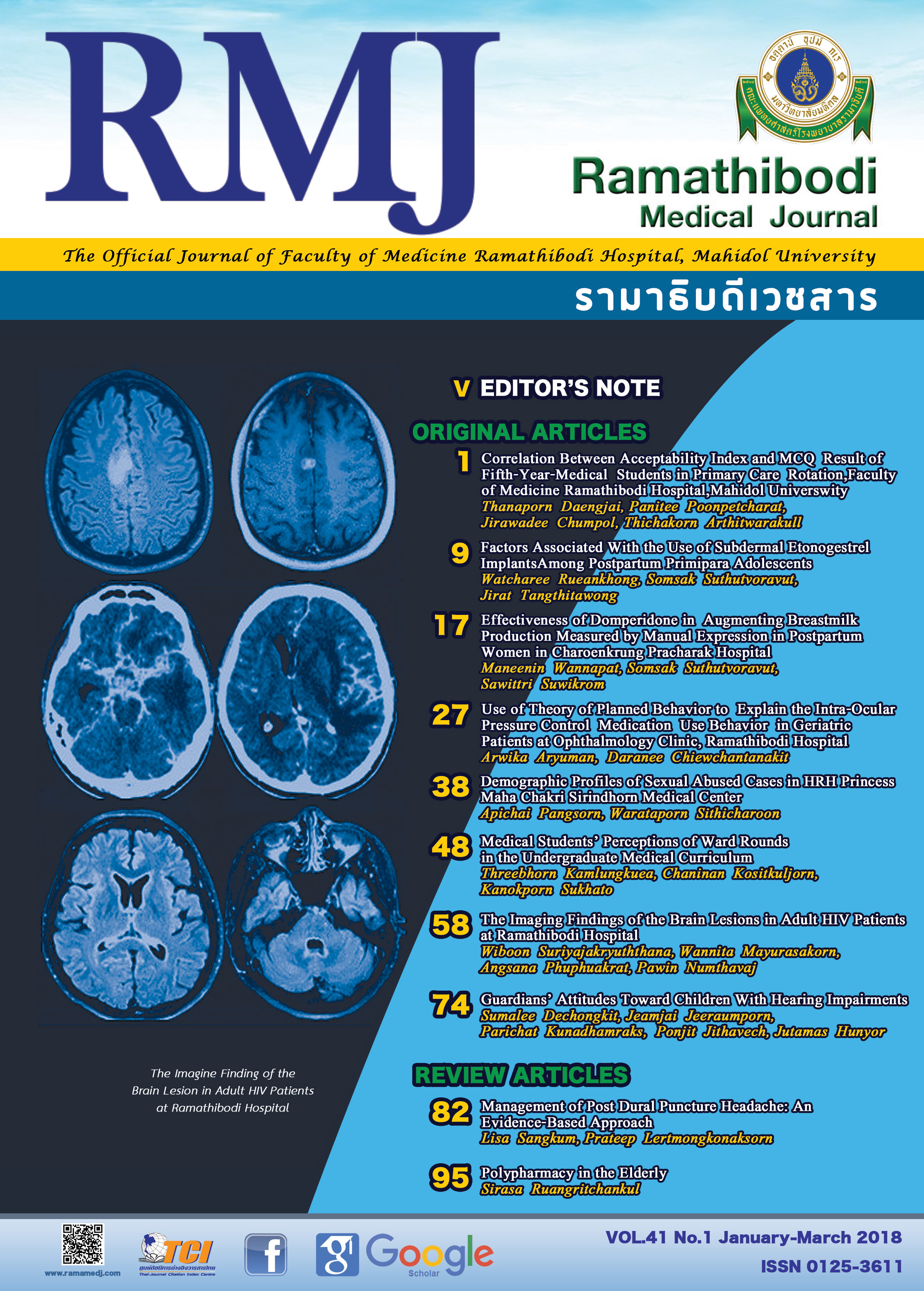
The Imaging Findings of the Brain Lesions in Adult HIV Patients at Ramathibodi Hospital
DOI:
https://doi.org/10.14456/rmj.2018.7Keywords:
CT, MRI, HIV, Immune reconstitution inflammatory syndrome, Highly active antiretroviral therapyAbstract
Background: The central nervous system (CNS) is major targets of HIV. Imaging plays major role for diagnosis. In the era of highly active antiretroviral therapy (HAART), immune reconstitution inflammatory syndrome (IRIS) frequently develops, causing worsening of opportunistic infection after initial of HAART and atypical imaging findings.
Objective: To evaluate radiographic findings of HIV-related brain lesions and imaging characteristics in CNS-IRIS at Ramathibodi Hospital.
Methods: Adult HIV patients who performed the first CT of the brain and first MRI of the brain were reviewed. The final diagnoses from medical records were assessed followed by CSF analysis, pathological report, and therapeutic treatment.
Results: Eighty-one HIV patients (64 CT brains and 44 MRI brains) with HIV-related brain lesions were diagnosed. There were 24.7% cryptococcal infection, 18.5% tuberculous infection, 8.6% HIV encephalopathy and progressive multifocal leukoencephalopathy (PML), 7.4% neurosyphilis, 6.2% toxoplasmosis and TB-IRIS, 2.5% primary CNS lymphoma, and others.
Conclusions: Cryptococcal infection was the most common disease of adult HIV-related brain lesions followed by tuberculous infection, HIV encephalopathy, and PML. Besides less basal meningitis and hydrocephalus in tuberculous meningitis and no demonstrated target lesion in tuberculoma compared with prior literatures, other imaging findings of each HIV-related brain lesions were not different from those of the prior studies.
References
Smith AB, Smirniotopoulos JG, Rushing EJ. From the archives of the AFIP: central nervous system infections associated with human immunodeficiency virus infection: radiologic-pathologic correlation. Radiographics. 2008;28(7):2033-2058. doi:10.1148/rg.287085135.
UNAIDS/WHO. Fact sheet: 2014 statistics. Geneva, Switzerland: World Health Organization; 2014.
Maschke M, Kastrup O, Esser S, Ross B, Hengge U, Hufnagel A. Incidence and prevalence of neurological disorders associated with HIV since the introduction of highly active antiretroviral therapy (HAART). J Neurol Neurosurg Psychiatry. 2000;69(3):376-380.
Masliah E, DeTeresa RM, Mallory ME, Hansen LA. Changes in pathological findings at autopsy in AIDS cases for the last 15 years. AIDS. 2000;14(1):69-74.
Burns DK, Risser RC, White CL. The neuropathology of human immunodeficiency virus infection. The Dallas, Texas, experience. Arch Pathol Lab Med. 1991;115(11):1112-1124.
Hongsakul K, Laothamatas J. Computer tomographic findings of the brain in HIV-patients at Ramathibodi Hospital. J Med Assoc Thai. 2008;91(6):895-907.
Post MJ, Thurnher MM, Clifford DB, et al. CNS-immune reconstitution inflammatory syndrome in the setting of HIV infection, part 1: overview and discussion of progressive multifocal leukoencephalopathy-immune reconstitution inflammatory syndrome and cryptococcal-immune reconstitution inflammatory syndrome. AJNR Am J Neuroradiol. 2013;34(7):1297-1307. doi:10.3174/ajnr.A3183.
Post MJ, Thurnher MM, Clifford DB, et al. CNS-immune reconstitution inflammatory syndrome in the setting of HIV infection, part 2: discussion of neuro-immune reconstitution inflammatory syndrome with and without other pathogens. AJNR Am J Neuroradiol. 2013;34(7):1308-1318. doi:10.3174/ajnr.A3184.
Shelburne SA, Hamill RJ, Rodriguez-Barradas MC, et al. Immune reconstitution inflammatory syndrome: emergence of a unique syndrome during highly active antiretroviral therapy. Medicine (Baltimore). 2002;81(3):213-227.
Robertson J, Meier M, Wall J, Ying J, Fichtenbaum CJ. Immune reconstitution syndrome in HIV: validating a case definition and identifying clinical predictors in persons initiating antiretroviral therapy. Clin Infect Dis. 2006;42(11):1639-1646.
Post MJD, Tate LG, Quencer RM et al. CT, MR, and pathology in HIV encephalitis and meningitis. AJR Am J Roentgenol. 1988;151:373-380.
Offiah CE, Naseer A. Spectrum of imaging appearances of intracranial cryptococcal infection in HIV/AIDS patients in the anti-retroviral therapy era. Clin Radiol. 2016;71(1):9-17. doi:10.1016/j.crad.2015.10.005.
Nelson CA, Zunt JR. Tuberculosis of the Central nervous system in immunocompromised patients: HIV infection and solid organ transplant recipients. Clin Infect Dis. 2011;53(9):915-926. doi:10.1093/cid/cir508.
Trivedi R, Saksena S, Gupta RK. Magnetic resonance imaging in central nervous system tuberculosis. Indian J Radiol Imaging. 2009;19(4):256-265. doi:10.4103/0971-3026.57205.
Smith MM, Andersona JC. Neurosyphilis as a cause of facial and vestibulocochlear nerve dysfunction: MR imaging features. AJNR Am J Neuroradiol. 2000;21(9):1673-1675.
Kumar GG, Mahadevan A, Guruprasad AS, et al. Eccentric target sign in cerebral toxoplasmosis: neuropathological correlate to the imaging feature. J Magn Reson Imaging. 2010;31(6):1469-1472. doi:10.1002/jmri.22192.
Mahadevan A, Ramalingaiah AH, Parthasarathy S, Nath A, Ranga U, Krishna SS. Neuropathological correlate of the "concentric target sign" in MRI of HIV-associated cerebral toxoplasmosis. J Magn Reson Imaging. 2013;38(2):488-495. doi:10.1002/jmri.24036.
Haldorsen IS, Espeland A, Larssonc EM. Central Nervous System Lymphoma: Characteristic Findings on Traditional and Advanced Imaging. AJNR Am J Neuroradiol. 2011;32(6):984-992. doi:10.3174/ajnr.A2171.
Dina TS. Primary central nervous system lymphoma versus toxoplasmosis in AIDS. Radiology. 1991;179(3):823-828.
Tan IL, McArthur JC, Venkatesan A, Nath A. Atypical manifestations and poor outcome of herpes simplex encephalitis in the immunocompromised. Neurology. 2012;79(21):2125-2132. doi:10.1212/WNL.0b013e3182752ceb.
Begley C, Amaraneni A, Lutwick L. Mycobacterium avium-intracellulare brain abscesses in an HIV-infected patient. IDCases. 2015;2(1):19-21.
Hariri OR, Minasian T, Quadri SA, et al. Histoplasmosis with deep CNS involvement: case presentation with discussion and literature review. J Neurol Surg Rep. 2015;76(1):e167-e172. doi:10.1055/s-0035-1554932.
Lindzen E, Jewells V, Bouldin T, Speer D, Royal W, Markovic-Plese S. Progressive tumefactive inflammatory central nervous system demyelinating disease in an acquired immunodeficiency syndrome patient treated with highly active antiretroviral therapy. J Neurovirol. 2008;14(6):569-573. doi:10.1080/13550280802304753.
Bahr N, Boulware DR, Marais S, Scriven J, Wilkinson RJ, Meintjes G. Central nervous system immune reconstitution inflammatory syndrome. Curr Infect Dis Rep. 2013;15(6):583-596. doi:10.1007/s11908-013-0378-5.


