
A Kaposi sarcoma-like presentation in Borderline Tuberculoid leprosy: a case report and review
Keywords:
Borderline Tuberculoid leprosy, Paucibacillary leprosy, Nested PCRAbstract
Leprosy, or Hansen’s disease, is a chronic infectious disease caused by Mycobacterium leprae. It affects the skin, peripheral nerves, and eyes1. Although curable with multidrug therapy (MDT), nearly 200,000 new cases are reported globally each year3. Early diagnosis and efforts to reduce social stigma are essential for disease control and prevention2.
Leprosy and human immunodeficiency virus (HIV) infection frequently coexist in endemic regions, yet their immunologic interaction remains complex and paradoxical. Although HIV-induced immunosuppression could theoretically increase susceptibility to Mycobacterium leprae, the clinical spectrum of leprosy is not markedly altered in most co-infected patients12,13.
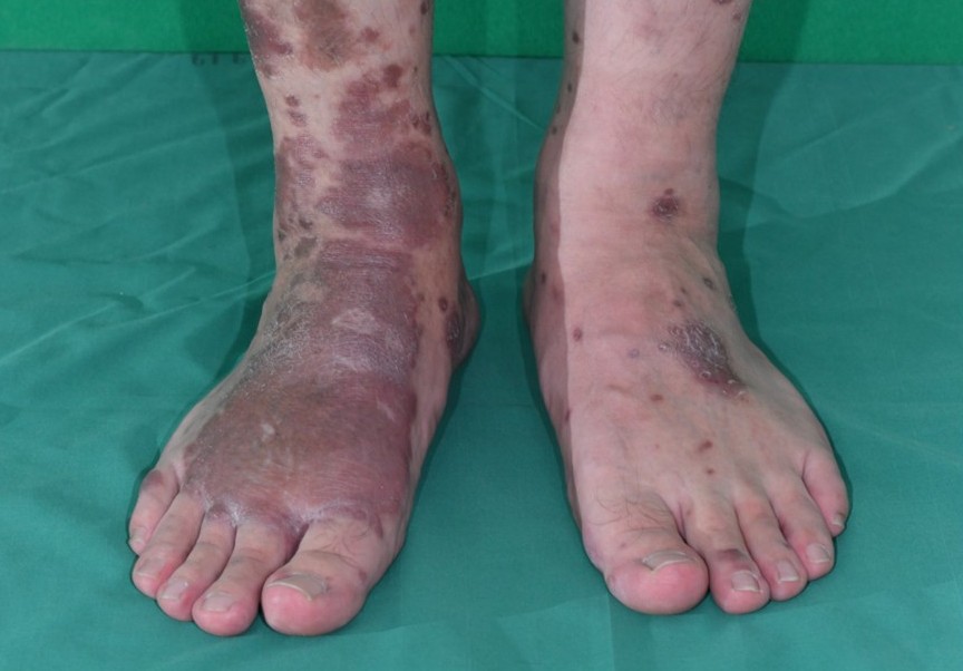
We present the case of a male patient with a known history of HIV, who presented with erythematous and violaceous lesions suggestive of Kaposi sarcoma. The lesions, characterized by both plaques and nodules, were widely distributed across the face, trunk, back, and extremities. A slit-skin smear yielded negative results, while a biopsy from the left arm revealed elongated sausage-like tuberculoid granulomas in the superficial perivascular plexus and along the neurovascular bundle. The Fite stain failed to reveal the organisms. Further examination using fluorescent staining with auramine O showed a positive result in the left ear. In addition, molecular analysis via Nested PCR targeting the M. leprae-specific repetitive element (RLEP) confirmed a diagnosis of leprosy. The patient underwent multidrug therapy, after which the skin lesions showed improvement. Fluorescent staining and nested PCR testing may aid in diagnosing cases of paucibacillary leprosy, where slit-skin smear examination is often inconclusive. These methods may serve as supplementary diagnostic tools, particularly in cases presenting with atypical skin lesions.
References
World Health Organization. Leprosy [Internet]. Geneva: World Health Organization; 2023 [cited 2025 May 19]. Available from: https://www.who.int/news-room/fact-sheets/detail/leprosy
Lockwood DN, Shetty V, Penna GO. Hazards of setting targets to eliminate disease: lessons from the leprosy elimination campaign. BMJ 2014;348:g1136.
Scollard DM, Adams LB, Gillis TP, Krahenbuhl JL, Truman RW, Williams DL. The continuing challenges of leprosy. Clin Microbiol Rev 2006;19:338-81.
Girma S, Avanzi C, Bobosha K, et al. Evaluation of Auramine O staining and conventional PCR for leprosy diagnosis: A comparative cross-sectional study from Ethiopia. PLoS Negl Trop Dis 2018;12:e0006706.
Martinez AN, Talhari C, Moraes MO, Talhari S. PCR-based techniques for leprosy diagnosis: from the laboratory to the clinic. PLoS Negl Trop Dis 2014;8:e2655.
Jiang H, Shi Y, Chokkakula S, et al. Utility of Multi-target Nested PCR and ELISPOT Assays for the Detection of Paucibacillary Leprosy: A Possible Conclusion of Clinical Laboratory Misdiagnosis. Front Cell Infect Microbiol 2022;12:814413.
Azevedo MC, Ramuno NM, Fachin LR, et al. qPCR detection of Mycobacterium leprae in biopsies and slit skin smear of different leprosy clinical forms. Braz J Infect Dis 2017;21:71-8.
Manandhar U, Adhikari R, Sayami G. Clinico-histopathological correlation of skin biopsies in leprosy. J Pathol Nep 2013;3:452-8.
Bathina A, Kollipara H, Sravani G, Laxmi VS. Beyond Classic Leprosy: Exploring Atypical Manifestations and Their Diagnostic Challenges. Cureus 2024;16:e75957.
Sawatkar GU, Sharma A, Keshavamurthy V, et al. Unusual clinical presentations in leprosy: a case series and review. Trans R Soc Trop Med Hyg 2023;117:489-95.
Mouchard A, Blaizot R, Graille J, Couppié P, Bertin C. Leprosy as immune reconstitution inflammatory syndrome in patients living with HIV: Description of French Guiana's cases over 20 years and systematic review of the literature. PLoS Negl Trop Dis 2022;16:e0010239.
Ustianowski AP, Lawn SD, Lockwood DN. Interactions between HIV infection and leprosy: a paradox. Lancet Infect Dis 2006;6:350-60.
Talhari C, Penna GO, Gonçalves HS, et al. Leprosy and HIV coinfection: a clinical, pathological, immunological, and therapeutic study of a cohort from a Brazilian referral center. J Infect Dis 2010;202:345–54.
Narang T, Bishnoi A, Dogra S, Saikia UN, Kavita. Alternate Anti-Leprosy Regimen for Multidrug Therapy Refractory Leprosy: A Retrospective Study from a Tertiary Care Center in North India. Am J Trop Med Hyg 2019;100:24-30.
Downloads
Published
How to Cite
Issue
Section
License
Copyright (c) 2026 Thai Journal of Dermatology

This work is licensed under a Creative Commons Attribution-NonCommercial-NoDerivatives 4.0 International License.
เนื้อหาและข้อมูลในบทความที่ลงตีพิมพ์ในวารสารโรคผิวหนัง ถือเป็นข้อคิดเห็นและความรับผิดชอบของผู้เขียนบทความโดยตรงซึ่งกองบรรณาธิการวารสาร ไม่จำเป็นต้องเห็นด้วย หรือร่วมรับผิดชอบใดๆ
บทความ ข้อมูล เนื้อหา รูปภาพ ฯลฯ ที่ได้รับการตีพิมพ์ในวารสารโรคผิวหนัง ถือเป็นลิขสิทธิ์ของวารสารฯ หากบุคคลหรือหน่วยงานใดต้องการนำทั้งหมดหรือส่วนหนึ่งส่วนใดไปเผยแพร่ต่อหรือเพื่อกระทำการใดๆ จะต้องได้รับอนุญาตเป็นลายลักอักษรจากบรรณาธิการวารสารโรคผิวหนังก่อนเท่านั้น

