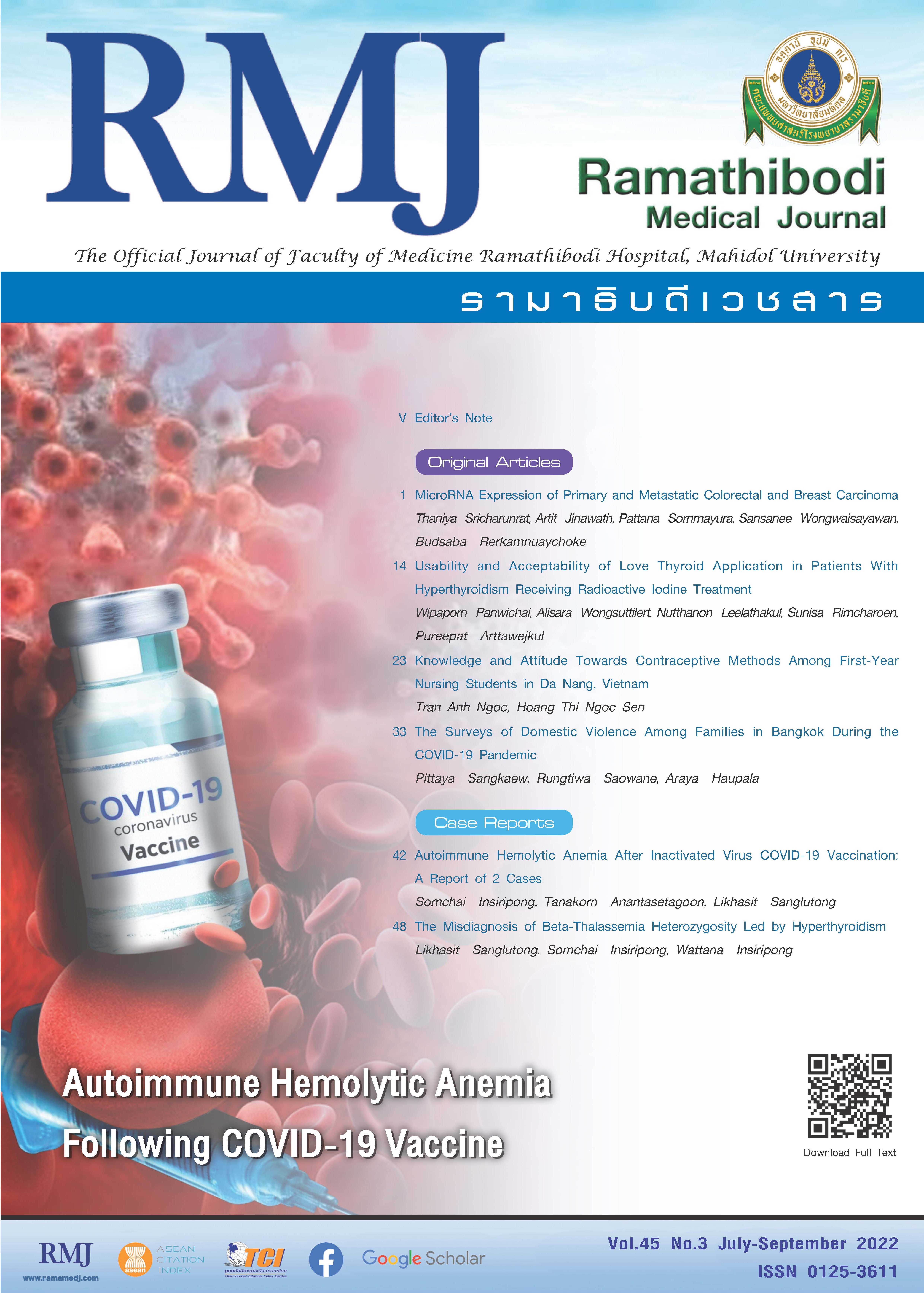
The Misdiagnosis of Beta-Thalassemia Heterozygosity Led by Hyperthyroidism
DOI:
https://doi.org/10.33165/rmj.2022.45.3.256975Keywords:
Hyperthyroidism , Fake Beta, Thalassemia, HeterozygosityAbstract
When patients have mild microcytic hypochromic anemia with slightly increased hemoglobin (Hb) A2 fraction, the most likely diagnosis is beta-thalassemia heterozygosity. But herein we found a patient who had all these hematological parameters but did not have beta-thalassemia heterozygosity. He was a 14-year-old Thai who presented with fatigue and heat intolerance for 2 weeks. His physical examination revealed mild diffuse enlargement of thyroid gland. Blood tests showed Hb 120 g/L, mean corpuscular volume (MCV) 72.1 fL, mean corpuscular hemoglobin (MCH) 23.3 pg/cell, free triiodothyronine (FT3) > 20 pg/mL, free thyroxine (FT4) > 5.0 ng/dL, thyrotropin < 2.5 mIU/L, serum ferritin 51.3 µg/L, Hb A2 3.8%. Besides primary hyperthyroidism, he was diagnosed with beta-thalassemia heterozygosity. After being treated with antithyroid drug for 6 months, his blood tests showed subclinical hyperthyroidism, Hb 146 g/L, MCV 83.3 fL, MCH 26.3 pg/cell, Hb A2 3.0%. Not only the thyroid hormones levels but also the Hb concentration, MCV, and the Hb A2 percentage became normal. Due to this inconsistency, the DNA analysis for beta-thalassemia genes was performed and found negative for numerous common and rare beta-thalassemia genes meanwhile beta-globin gene sequencing appeared normal. It should be concluded that hyperthyroidism could induce slightly elevated Hb A2 percentage and mild hypochromic microcytic anemia in a normal individual, leading to the misdiagnosis of beta-thalassemia heterozygosity. In other words, Hb analysis should not be performed during hyperthyroidism and it should be delayed till achievement of the euthyroid stage.
References
Zhang J, He J, Mao X, et al. Haematological and electrophoretic characterisation of β-thalassaemia in Yunnan province of Southwestern China. BMJ Open. 2017;7(1):e013367. doi:10.1136/bmjopen-2016-013367
Gu H, Wang YX, Du MX, Xu SS, Zhou BY, Li MZ. Effectiveness of using mean corpuscular volume and mean corpuscular hemoglobin for beta-thalassemia carrier screening in the Guangdong population of China. Biomed Environ Sci. 2021;34(8):667-671. doi:10.3967/bes2021.094
Ou Z, Li Q, Liu W, Sun X. Elevated hemoglobin A2 as a marker for β-thalassemia trait in pregnant women. Tohoku J Exp Med. 2011;223(3):223-226. doi:10.1620/tjem.223.223
Verma S, Gupta R, Kudesia M, Mathur A, Krishan G, Singh S. Coexisting iron deficiency anemia and beta thalassemia trait: effect of iron therapy on red cell parameters and hemoglobin subtypes. ISRN Hematol. 2014;2014:293216. doi:10.1155/2014/293216
Kravets I. Hyperthyroidism: diagnosis and treatment. Am Fam Physician. 2016;93(5):363-370.
Johnson-Wimbley TD, Graham DY. Diagnosis and management of iron deficiency anemia in the 21st century. Therap Adv Gastroenterol. 2011;4(3):177-184. doi:10.1177/1756283X11398736
Ebert BL, Mak R, Pretz JL, et al. High throughput screen to identify small molecules that differentially regulate expression of the globin genes. Blood. 2005;106(11):3632. doi:10.1182/blood.V106.11.3632.3632
Jaafer FH, Al-Tememi WF, Ali HH. The influence of thyroid status on hemoglobin A2, F expression. New Iraqi J Med. 2009;5(3):78-81. Accessed March 27, 2022. https://applications.emro.who.int/imemrf/New_Iraqi_J_Med/New_Iraqi_J_Med_2009_5_3_78_81.pdf
Akasheh MS. Graves’ disease mimicking beta-thalassemia trait. Postgrad Med J. 1994;70(822):300-301. doi:10.1136/pgmj.70.822.300
Al-Amodi AM, Ghanem NZ, Aldakeel SA, et al. Hemoglobin A2 (HbA2) has a measure of unreliability in diagnosing β-thalassemia trait (β-TT). Curr Med Res Opin. 2018;34(5):945-951. doi:10.1080/03007995.2018.1435520
Ahmed SS, Mohammed AA. Effects of thyroid dysfunction on hematological parameters: case controlled study. Ann Med Surg (Lond). 2020;57:52-55. doi:10.1016/j.amsu.2020.07.008
Sharma P, Das R, Trehan A, et al. Impact of iron deficiency on hemoglobin A2% in obligate β-thalassemia heterozygotes. Int J Lab Hematol. 2015;37(1):105-111. doi:10.1111/ijlh.12246
Maheshwari KU, Rajagopalan B, Samuel TR. Variations in hematological indices in patients with of thyroid dysfunction. Int J Contemp Med Res. 2020;7(1):A5-A7. doi:10.21276/ijcmr.2020.7.1.9
Dorgalaleh A, Mahmoodi M, Varmaghani B, et al. Effect of thyroid dysfunctions on blood cell count and red blood cell indice. Iran J Ped Hematol Oncol. 2013;3(2):73-77.
Hegazi MO, Ahmed S. Atypical clinical manifestations of graves’ disease: an analysis in depth. J Thyroid Res. 2012;2012:768019. doi:10.1155/2012/768019
Gianoukakis AG, Gupta S, Tran TN, Richards P, Yehuda M, Tomassetti SE. Graves' disease patients with iron deficiency anemia: serologic evidence of co-existent autoimmune gastritis. Am J Blood Res. 2021;11(3):238-247.
Zhang Y, Xue Y, Cao C, et al. Thyroid hormone regulates hematopoiesis via the TR-KLF9 axis. Blood. 2017;130(20):2161-2170. doi:10.1182/blood-2017-05-783043
Ma Y, Freitag P, Zhou J, Brüne B, Frede S, Fandrey J. Thyroid hormone induces erythropoietin gene expression through augmented accumulation of hypoxia-inducible factor-1. Am J Physiol Regul Integr Comp Physiol. 2004;287(3):R600-R607. doi:10.1152/ajpregu.00115.2004
Valencia ME, Kyaw Y, Chinnasamy E. Hyperthyroidism as an under-recognized reversible cause of microcytosis. Endocrine Abstracts. 2021;74:NCC17. doi:10.10.1530/endoabs.74.NCC17
Brining L, Cadre RR. Graves’ disease with leukopenia and microcytosis. In: Proceedings of UCLA Healthcare Volume 19. Department of Medicine - UCLA; 2015. Accessed March 27, 2022. https://proceedings.med.ucla.edu/wp-content/uploads/2016/11/Graves%E2%80%99-Disease-with-Leukopenia-and-Microcytosis.pdf
Blanc L, Papoin J, Vidal M, et al. Ineffective erythropoiesis is the major cause of microcytic anemia in the TSAP6/Steap3 null mouse model. Blood. 2014;124(21):1332. doi:10.1182/blood.V124.21.1332.1332
Gil-Morales C, Costa M, Tennant K, Hibbert A. Incidence of microcytosis in hyperthyroid cats referred for radioiodine treatment. J Feline Med Surg. 2021;23(10):928-935. doi:10.1177/1098612X20983973
Downloads
Published
How to Cite
Issue
Section
License
Copyright (c) 2022 Ramathibodi Medical Journal

This work is licensed under a Creative Commons Attribution-NonCommercial-NoDerivatives 4.0 International License.


