
Medical Modelling Design and Production of Ultrasound-Guided Internal Jugular Vein for Residency Training Medical Modelling Design
Article Sidebar
Main Article Content
Abstract
Objective: To develop an ultrasound-guided internal jugular vein model for residency training
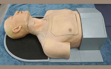
Materials and Methods: The researcher invited seven medical professionals within the Faculty of Medicine Siriraj Hospital who had experience in ultrasound-guided internal jugular vein procedure with real patients and had at least 3 years of teaching and training as resident physicians, aged between 35 - 60 years, to test the performance and features of an ultrasound-guided internal jugular vein model. The objective was to test the properties of silicone rubber suitable for viewing ultrasound images before producing a mannequin. This model consisted of interesting three parts: subcutaneous fat, subcutaneous silicone rubber, skin, and blood vessels. The properties and performance of the model were tested according to the procedure using ultrasound-guided internal venous catheter insertion in the jugular, and a satisfaction assessment form from seven medical specialists was used to evaluate the model.
Results: All seven medical professionals tested the properties of silicone rubber and found that silicone rubber E is suitable for replacing the subcutaneous fat layer, silicone rubber F is suitable for replacing the skin, and J silicone rubber is suitable for replacing blood vessels. The test of the model's properties indicated a level of satisfaction at the "very satisfied" level. The evaluation of the model's effectiveness in the ultrasound-guided internal venous catheter insertion procedure was at the level of satisfaction, ranging from "good".
Conclusion: The researcher has successfully developed a qualified ultrasound-guided internal jugular vein model that can be effectively used for teaching and learning purposes.
Article Details

This work is licensed under a Creative Commons Attribution-NonCommercial-NoDerivatives 4.0 International License.


References
Rivera AM, Strauss KW, van Zundert AA, Mortier EP. Matching the peripheral intravenous catheter to the in- dividual patient. Acta Anaesthesiol Belg 2007;58:19-25.
Feldman R. Central venous access. In: Richman E,Simon M, editors. Emergency medicine procedure.United States: McGrawHill;2002.
Paula Ferrada. How To Do Internal Jugular Vein Cannulation, Ultrasound-Guided 2020 Available from: https://www.merckmanuals.com/professional/critical-care-medicine/how-to-do-central-vascular-procedures/how-to-do-internal-jugular-vein-cannulation,-ultrasound-guided.
Joshua Kolikof, Katherine Peterson, Annalee M. Baker. Central Venous Catheter. StatPearls. 2022.
อลิสสรา วณิชกุลบดี. บทบาทการใช้คลื่นเสียงความถี่สูงในห้องฉุกเฉิน.วชิรเวชสารและวารสารเวชศาสตร์เขตเมือง. 2016;60(4):297-308.
Young, M. P., Yuo, T. H., Manaker, S., & Hall, K. K. Overview of complications of central venous catheters and their prevention in adults.
KARAKITSOS, Dimitrios, et al. Real-time ultrasound-guided catheterisation of the internal jugular vein: a prospective comparison with the landmark technique in critical care patients. Critical Care, 2006, 10.6:1-8.
Schofer JM, Nomura JT, Bauman MJ, Sierzenski PR. Prospective durability testing of a vascular access phantom. The Western Journal of Emergency Medicine. 2010;11(4):302-305.
ÁLVAREZ, JM López, et al. Revision of Training Models on Ultrasound-Guided Vascular Access. Presentation of an Animal Model. Ultrasound Imaging: Current Topics, 2022, 117.
ยาใจ อภิบุณโยภาส. การใส่สายสวนหลอดเลือดดำส่วนกลาง. ธรรมศาสตร์เวชสาร 2557. 14;1:79-92.
Margaret Lin-Martore MD, Shruti Kant MBBS, Bridget C. O’Brien Ph.D. Procedural skill maintenance: Perspectives and motivations of pediatric emergency medicine faculty. AEM Education and Training 2021;5:4.
Phrampus PE. Simulation and integration into patient safety systems. Simul Healthcare. 2018;13(4):225–226.
EnvironMolds. What is Condensation Cure and Addition Cure Silicone? 2022 [cited 2022 Oct 1]. Available from: https://www.artmolds.com/condensation-addition-cured-silicone
Phadungsak Silakorn, M.sc., Wangcha Chumthap, BSc., Somchai Chongpipatchaiporn, B.Sc., Arsada Khunvut, B.Sc., Supenya Vsrothai, M.D. : Silicone Rubber Development for Medical Model Production. Siriraj Med J 2015; 67: 123-127.
Brick In The Yard Mold Supply. [cited 2018 February 11]. RTV Silicone Tutorial: Shore A Scale [Video file]. Available from: https://www.youtube.com/watch?v=Oj9fwv6BcUM
กรวีร์ เทพสัมฤทธิ์พร. Acute Venous Catheterization in General Practice. ม.ป.ป. 499-505.
LEE, Jeong Gil, et al. Effect of Trendelenburg position on right and left internal jugular vein cross-sectional area. Korean Journal of Anesthesiology, 2014, 67.5:305-309.
JUDICKAS, Šarūnas, et al. Is the Trendelenburg position the only way to better visualize internal jugular veins?. ACTA Medica Lituanica, 2018, 25.3:125.
