
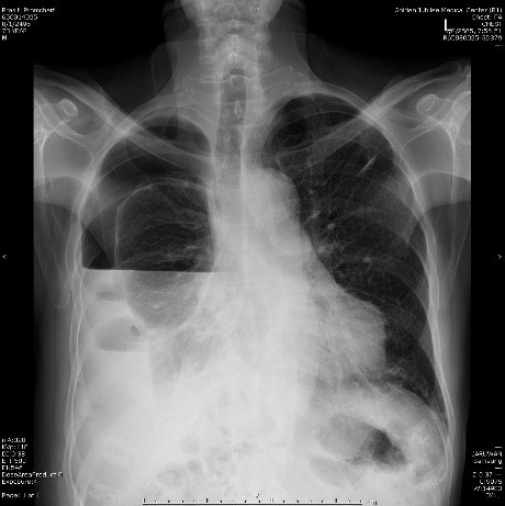
Entrance Skin Dose from Chest X-Ray Radiograph at Radiology Division, Golden Jubilee Medical Center
Article Sidebar
Main Article Content
Abstract
Objective: To study the entrance skin dose (ESD) of patients underwent chest x-ray radiograph at radiology division, Golden Jubilee Medical Center, Faculty of medicine Siriraj hospital, Mahidol University, and compare with the diagnostic reference level of the International Atomic Energy Agency and other studies.
Materials & Methods: This study is an observational descriptive study. The patients aged 18-70 years were selected, with body weight ranging from 60±15 kg. underwent with digital radiography system, Samsung XGEO GC80 model. Demographic data and technical parameters including gender, age, weight, height, chest thickness, kilovoltage peak, milli-ampere*second were collected for 304 patients during October 2021 – May 2022. Estimate ESD was calculated from vender’s formula. The median and 3rd quartile of the calculated dose was used to compare with the DRLs of national, international and other relevant studies.
Results: The results revealed that the median and 3rd quartile of ESD were 0.13 mGy and 0.15 mGy, respectively. The means of milli-ampere*second for AEC and manual mAs were 2.89 and 3.43 mAs, respectively. Both techniques use the same 110 kVp and mAs ranges 1.9 – 6 depending on technique and patient’s thickness.
Conclusion: ESD from chest radiograph obtained was below the DRL from IAEA and other corresponding studies. This study led to the development for optimization proper kVp and mAs parameters to safety level of radiation. However, image quality must be adequate to provide the information required for the clinical diagnosis.
Article Details

This work is licensed under a Creative Commons Attribution-NonCommercial-NoDerivatives 4.0 International License.


References
กรมวิทยาศาสตร์การแพทย์ กระทรวงสาธารณสุข. National Diagnostic Reference Levels in Thailand 2021 (ค่าปริมาณรังสีอ้างอิงในการถ่ายภาพรังสีวินิจฉัยทางการแพทย์ของประเทศไทย 2564). บริษัท บียอนด์ พับลิสชิ่ง จำกัด, 2564.
ประภัสสร ไกรหาญ, ดวงฤดี สุภมาตย์. การประเมินค่าดัชนีชี้วัดปริมาณรังสี (Exposure index:EI, Deviation index :DI) ในการถ่zยภาพทรวงอกเด็กอายุ 0-12 ปี โรงพยาบาลสิรินธร จังหวัดขอนแก่น. วารสารสุขภาพและสิ่งแวดล้อมศึกษา 2564;6(4):18-27.
International Atomic Energy Agency. International basic safety standard for protection against ionizing radiation and for the safety of radiation source, IAEA Safety Series No. 115. Veinna: International Atomic Energy Agency, 1996.
สำนักรังสี และเครื่องมือแพทย์ กรมวิทยาศาสตร์การแพทย์. ปริมาณรังสีอ้างอิงการถ่ายภาพรังสีวินิจฉัยด้วยเครื่องเอกซเรย์ทั่วไป [อินเทอร์เน็ต]. [เข้าถึงเมื่อ 15 มิถุนายน 2565]. เข้าถึงได้จาก http://brmd.dmsc.moph.go.th/radiation/
ศิริวรรณ บุญชรัตน์, ภัทรพงศ์ เหมัษฐิติ. ค่าปริมาณรังสีอ้างอิง จากการถ่ายภาพรังสีวินิจฉัย ด้วยเครื่องเอกซเรย์ทั่วไป (Diagnostic reference levels in General radiography) สำนักรังสีและ เครื่องมือแพทย์.วารสารวิชาการสาธารณสุข 2559;25(4):632-40.
ลัดดา เย็นศรี. การศึกษาเปรียบเทียบปริมาณรังสีที่ผู้ป่วยได้รับ จากการถ่ายภาพรังสีทรวงอกด้วยระบบ CR และ DR. วารสารเครือข่ายวิทยาลัยพยาบาลและการสาธารณสุขภาคใต้ 2559;3(1):129–39.
ไพรัตน์ มุณี, วิชุดา สิริเมธาธโนปกรณ์, ปรียานุช มโนธรรม. การศึกษาระดับรังสีอ้างอิงสำหรับการถ่ายภาพรังสีดิจิทัล: กรณีศึกษาโรงพยาบาลศิริราช. วารสารรังสีวิทยาศิริราช 2561;5(1):25-32.
Promduang A., Pongnapang, N., Ritlumlert, N., Tangruangkiat, T., Phonlakrai, M. (2019). A Study of Entrance Surface Air Kerma for Patients Undergoing Chest and Abdomen from Digital Radiography at Chulabhorn Hospital. Journal of Health Science and Medical Research. 37(1):51-60.
จริญญา เต็งชัยภูมิ, จินดาวัลย์ วิบูลย์อุทัย, เพชรากร หาญพานิชย์. การศึกษาความสัมพันธ์ของปริมาณรังสีที่เหมาะสมตามเกณฑ์มาตรฐาน และเทคนิคการถ่ายภาพทางรังสี ในโรงพยาบาลหนองสูง จังหวัดมุกดาหาร. วารสารการแพทย์และวิทยาศาสตร์สุขภาพ 2563;27:111-22.
ยุทธนา เนตรวงศ์. การประเมินปริมาณรังสีที่ผิวผู้ป่วยจากการถ่ายภาพรังสีวินิจฉัยทั่วไป ในสถาบันประสาทวิทยาโดยใช้ค่าผลคูณปริมาณรังสีกับพื้นที่. วารสารกรมการแพทย์ 2563;45(3):90-7.
ต้องจิต มหาจันทวงศ์, ธวัชชัย ปราบศัตรู, วรนันท์ คีรีสัตยกุล, สมศักดิ์ วงษาศานนท์, วราภรณ์ สุดใจ. การประเมินปริมาณรังสีที่ผิวที่ผู้ป่วยได้รับในการถ่ายภาพทางรังสีดิจิทัล ในโรงพยาบาลศรีนครินทร์. วารสารศรีนครินทร์เวชสาร 2564;36(1):31-8.
Hart, D., Hillier, M. C. and Shrimpton, P. C. Doses to Patients from Radiographic and Fluoroscopic X-ray Imaging Procedures in the UK – 2010 Review. Health Protection Agency (UK) - HPA-CRCE-034. (2012).
Yonekura, Y. Japan Network for Research and Information on Medical Exposure (J-RIME). Diagnostic reference levels based on latest surveys in Japan – Japan DRLs – 2015. (2015). Available on http://www.radher.jp/J-RIME/report/DRLhoukoku syoEng.pdf (cited 2022.6.18).
Vañó E, Miller DL, Martin CJ, Rehani MM, Kang K, Rosenstein M, Ortiz-López P, Mattsson S, Padovani R, Rogers A; Authors on behalf of ICRP. ICRP Publication 135: Diagnostic Reference Levels in Medical Imaging. Ann ICRP. 2017 Oct;46(1):1-144.
