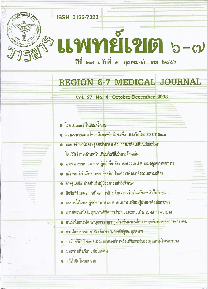
ความสัมพันธ์ระหว่างความหนาของกะโหลกศีรษะที่วัดโดยตรงด้วยเครื่องวัดกับที่วัดโดยภาพถ่ายรังสีของกะโหลกศีรษะสามมิติ
บทคัดย่อ
This is a descriptive study that measure the thickness of parietal bone in Thai adult cadavers and find out its correlation to three-dimensional CT scan. A total of 65 (male 34 and female 31) cadaveric skulls were used in this study. The calvarial thickness is measured in 9 points on each parietal area of skull by Depth micrometer series 128-101 (Mitutoyo Corporation, Kanagawa, Japan). The three-dimensional CT scans were made on a GE light speed VCT 64 scanner (General Electrics Medical System, Milwaukee, Wisconsin). Mean thickness of all parietal bones was 6.68 ± 1.94 mm (0.84-15.59 mm). At point 5, mean thickness measured by micrometer was 7.13 1 ± 2.28 mm and 7.00 ± 2.22 mm with three-dimensional CT scan, respectively. Mean difference was 0.13 mm that statistically significant (p-value < 0.01) but the upper limit of 95% confidence interval of all difference was only 0.28 mm that not clinically significant. The relationship between the two measurement modalities could had equation of relationship as micrometer = 0.025 + 1.025 (3D-CT). The study concluded that agreement between the micrometer and three-dimensional CT measurements was acceptable.
ดาวน์โหลด
เผยแพร่แล้ว
รูปแบบการอ้างอิง
ฉบับ
ประเภทบทความ
สัญญาอนุญาต
ลิขสิทธิ์บทความเป็นของผู้เขียนบทความ แต่หากผลงานของท่านได้รับการพิจารณาตีพิมพ์ลงวารสารแพทย์เขต 4-5 จะคงไว้ซึ่งสิทธิ์ในการตีพิมพ์ครั้งแรกด้วยเหตุที่บทความจะปรากฎในวารสารที่เข้าถึงได้ จึงอนุญาตให้นำบทความในวารสารไปใช้ประโยชน์ได้ในเชิงวิชาการโดยจำเป็นต้องมีการอ้างอิงถึงชื่อวารสารอย่างถูกต้อง แต่ไม่อนุญาตให้นำไปใช้ในเชิงพาณิชย์


