โคนบีมซีที: ประโยชนที่ได้รับกับโอกาสเกิดโรคมะเร็งในยุคทันตกรรมดิจิตอล
โคนบีมซีทีกับการเกิดโรคมะเร็ง
คำสำคัญ:
โคนบีมซีที, รังสีวินิจฉัย, ทันตกรรมดิจิตอล, มะเร็งศีรษะและลําคอบทคัดย่อ
ในยุคทันตกรรมดิจิตอลการตรวจช่องปากด้วยเครื่องถ่ายภาพโคนบีมซีที (CBCT) เป็นอุปกรณ์ที่ถูกใช้อย่างแพร่หลายและมีราคาย่อมเยามากขึ้น เป็นหนึ่งในวิธีการตรวจที่เป็นมาตรฐานของการวินิจฉัย การรักษา จนถึงการติดตามอาการอย่างไรก็ตามหากไม่มีการควบคุมการใช้งาน และการเรียนรู้ถึงผลกระทบของการใช้งานเกินขอบเขตหรือเกินความจําเป็นย่อมทําให้เกิดผลเสียต่อผู้ป่วยได้ โดยเฉพาะอย่างยิ่งในแง่ของการเพิ่มความเสี่ยงในการเกิดโรคมะเร็ง บทความนี้จึงรวบรวมข้อมูลตั้งแต่ความรู้เบื้องต้นเกี่ยวกับรังสีและผลของร่างกายต่อการรับรังสีเอ็กซ์ วิธีการวัดค่ารังสี การป้องกันรังสีไม่ให้รับเกินความจําเป็น แต่ยังเพียงพอต่อการวินิจฉัยและสมเหตุสมผล รวมไปถึงการควบคุมปริมาณรังสี โดยเลือกค่าพารามิเตอร์ตามแต่ละสถานการณ์ ไม่ควรใช้ค่าเดียวในทุกสถานการณ์ โดยเฉพาะอย่างยิ่งในเด็กและสตรีที่มีโอกาสสะสมรังสีมากกว่า โดยยึดหลัก 3 ประการของการสัมผัสรังสี จากคณะกรรมาธิการระหว่างประเทศด้านการป้องกันรังสี รวมทั้งมีการอธิบายทําความเข้าใจแก่ผู้ป่วยถึงโอกาสการเกิดมะเร็งจากรังสีวินิจฉัยแม้จะน้อยมากก็ตาม และการขอความยินยอมทุกครั้งก่อนผู้ป่วยจะสัมผัสรังสี
เอกสารอ้างอิง
Scarfe WC, Farman AG. What is cone-beam CT and how does it work? Dent Clin North Am 2008; 52(4): 707-30.
Silva MA, Wolf U, Heinicke F, Bumann A, Visser H, Hirsch E. Cone-beam computed tomography for routine orthodontic treatment planning: a radiation dose evaluation. Am J Orthod Dentofacial Orthop 2008; 133(5): 640.e1-5.
Scarfe WC, Farman AG. Cone beam computed tomography: a paradigm shift for clinical dentistry. Aust Dent Pract 2008; 19: 102-6.
Miracle AC, Mukherji SK. Conebeam CT of the head and neck, part 1: physical principles. AJNR Am J Neuroradiol 2009; 30(6): 1088-95.
Kamburoglu K, Murat S, Kilic C. Cone beam computed tomography in dentistry: a review. Clinics 2011; 66(6): 1073-81.
De Vos W, Casselman J, Swennen GRJ. Cone-beam computerized tomography (CBCT) imaging of the oral and maxillofacial region: A systematic review of the literature. Int J Oral Maxillofac Surg 2009; 38(6): 609-25.
Misch CE. Contemporary implant dentistry. 3rd ed. Mosby: St. Louis; 2011.
Kapila S, Conley RS, Harrell WE Jr. The current status of cone beam computed tomography imaging in orthodontics. Dentomaxillofac Radiol 2011; 40(1): 24-34.
Patel S, Horner K, Wilson R. The use of cone beam computed tomography in endodontics. Int Endod J 2009; 42(9): 755-73.
Loubele M, Maes F, Jacobs R, Schutyser F, Suetens P. Comparative localized bone qualityassessment based on cone-beam computed tomography. Clin Oral Implants Res 2009; 20(4): 520-5.
Vandenberghe B, Jacobs R, Bosmans H. Modern dental imaging: A review of the current technology and clinical applications in dental practice. Eur Radiol 2010; 20(11): 2637-55.
Tyndall DA, Rathore S. Cone-beam CT diagnostic applications: Caries, periodontal bone assessment, and endodontic applications. Dent Clin North Am 2008; 52(4): 825-41.
Pauwels R, Beinsberger J, Collaert B, Theodorakou C, Rogers J, Walker A, et al. Effective dose range for dental cone beam computed tomography scanners. Eur J Radiol 2012; 81(2): 267-71.
Dunne-Daly CF. Principles of radiotherapy. Br J Hosp Med (Lond) 1999; 74: C166-9.
Borrego-Soto G, Ortiz-López R, Rojas-Martínez A. Ionizing radiation-induced DNA injury and damage detection in patients with breast cancer. Genet Mol Biol 2015; 38(4): 420-32.
International Commission on Radiological Protection (ICRP). ICRP Publication 152. Annals of the ICRP 2022; 51(3): 1-3.
Cardis E, Howe G, Ron E, Bebeshko V, Bogdanova T, Bouville A, et al. Cancer consequences of the Chernobyl accident: 20 years on. J Radiol Prot 2006; 26: 127-40.
Food and Drug Administration. What are the radiation risks from CT? [online]. 2017; [cited 2022 Nov 4]: [4 screens]. Available from: URL: https://www.fda.gov/radiation-emitting-products/medical-x-ray-imaging/what-are-radiation-risks-ct.
International Agency for Research on Cancer. IARC monographs on the evaluation of carcinogenic risks to humans volume 75, part 1: X- and gamma -radiation, and neutrons. [online]. 2000; [cited 2022 Nov 4]. Available from: https://publications.iarc.fr/93.
Linet MS, Slovis TL, Miller DL, Kleinerman R, Lee C, Rajaraman P, et al. Cancer risks associated with external radiation from diagnostic imaging procedures. CA Cancer J Clin 2012; 62: 75-100.
Claus EB, Calvocoressi L, Bondy ML, Schildkraut JM, Wiemels JL, Wrensch M. Dental x-rays and risk of meningioma. Cancer 2012; 118(18): 4530-7.
Hall EJ, Giaccia AJ. Radiobiology for the radiologist. 7th ed. Philadelphia: Lippincott Williams & Wilkins; 2012.
U.S. Environmental Protection Agency. Radiation: health effects. [online]. 2023; [cited 2024 Nov 27]. Available from: URL: https://www.epa.gov/radiation/radiation-health-effects.
International Commission on Radiological Protection (ICRP). The 2007 recommendations of the international commission on radiological protection. ICRP publication 103. Ann ICRP 2007; 37(2-4): 1-332.
Al Najjar A, Colosi D, Dauer LT, Prins R, Patchell G, Branets I, et al. Comparison of adult and child radiation equivalent doses from 2 dental cone-beam computed tomography units. Am J Orthod Dentofac Orthop 2013; 143(6): 784-92.
Ludlow JB, Ivanovic M. Comparative dosimetry of dental CBCT devices and 64-slice CT for oral and maxillofacial radiology. Oral Surg Oral Med Oral Pathol Oral Radiol Endod 2008; 106(1): 106-14.
Pauwels R, Beinsberger J, Collaert B, Theodorakou C, Rogers J, Walker A, et al. Effective dose range for dental cone beam computed tomography scanners. Eur J Radiol 2012; 81(2): 267–71.
Ludlow JB, Walker C. Assessment of phantom dosimetry and image quality of i-CAT FLX cone-beam computed tomography. Am J Orthod Dentofac Orthop 2013; 144(6): 802-17.
Al-Okshi A, Nilsson M, Petersson A, Wiese M, Lindh C. Using GafChromic film to estimate the effective dose from dental cone beam CT and panoramic radiography. Dentomaxillofac Radiol 2013; 42(7): 20120343. (8 pages).
Koivisto J, Kiljunen T, Tapiovaara M, Wolff J, Kortesniemi M. Assessment of radiation exposure in dental cone-beam computerized tomography with the use of metal-oxide semiconductor field-effect transistor (MOSFET) dosimeters and Monte Carlo simulations. Oral Surg Oral Med Oral Pathol Oral Radiol 2012; 114(3): 393-400.
Lee C, Lee SS, Kim JE, Huh KH, Yi WJ, Heo MS, et al. Comparison of dosimetry methods for panoramic radiography: thermoluminescent dosimeter measurement versus personal computer–based Monte Carlo method calculation. Oral Surg Oral Med Oral Pathol Oral Radiol 2016; 121(3): 322-9.
Servomaa A, Tapiovaara M. Organ dose calculation in medical x-ray examinations by the program PCXMC. Radiat Prot Dosim 1998; 80(1-3): 213-9.
Khelassi-Toutaoui N, Berkani Y, Tsapaki V, Toutaoui AE, Merad A, Frahi-Amroun A, et al. Experimental evaluation of PCXMC and prepare codes used in conventional radiology. Radiat Prot Dosim 2008; 131(3): 374-8.
Tapiovaara M, Lakkisto M, Servomaa A. A PC-based Monte Carlo program for calculating patient doses in medical x-ray examinations. In: Report STUK-A139. Helsinki, Finland: Finnish Centre for Radiation and Nuclear Safety; 1997. p. 5-25.
Tapiovaara M. PCXMC 2.0 supplementary programs user’s guide. Helsinki, Finland: STUK; 2012.
Ludlow JB, Davies-Ludlow LE, Brooks SL. Dosimetry of two extraoral direct digital imaging devices: NewTom cone beam CT and Orthophos Plus DS panoramic unit. Dentomaxillofac Radiol 2003; 32: 229-34.
Arai Y, Tammisalo E, Iwai K, Hashimoto K, Shinoda K. Development of a compact computed tomographic apparatus for dental use. Dentomaxillofac Radiol 1999; 28: 245-8.
Scarfe WC, Farman AG. What is cone-beam CT and how does it work? Dent Clin North Am 2008; 52: 707-30.
Kadesjö N, Lynds R, Nilsson M, Shi XQ. Radiation dose from X-ray examinations of impacted canines: cone beam CT vs two-dimensional imaging. Dentomaxillofac Radiol 2018; 47(3): 20170305. (9 pages).
Roberts J, Drage N, Davies J, Thomas D. Effective dose from cone beam CT examinations in dentistry. Br J Radiol 2009; 82(973): 35-40.
Lorenzoni DC, Bolognese AM, Garib DG, Guedes FR, Sant'anna EF. Cone-beam computed tomography and radiographs in dentistry: aspects related to radiation dose. Int J Dent 2012; 2012: 813768. (10 pages).
Ludlow J, Davies-Ludlow L, Brooks S. Dosimetry of two extraoral direct digital imaging devices: NewTom cone beam CT and Orthophos plus DS panoramic unit. Dentomaxillofac Radiol 2003; 32(4): 229-34.
Garcia Silva MA, Wolf U, Heinicke F, Gründler K, Visser H, Hirsch E. Effective dosages for recording Veraviewepocs dental panoramic images: analog film, digital, and panoramic scout for CBCT. Oral Surg Oral Med Oral Pathol Oral Radiol Endod 2008; 106(4): 571-77.
Chinem LA, Vilella Bde S, Maurício CL, Canevaro LV, Deluiz LF, Vilella Ode V. Digital orthodontic radiographic set versus cone-beam computed tomography: an evaluation of the effective dose. Dent Press J Orthod 2016; 21(4): 66-72.
Sedentexct. Brussels: Sedentexct 2020-2021. [online]. 2021; [cited 2024 Nov 27]. Available from: URL: https://sedentexct.eu/content/basicprinciples-use-dental-cone-beam-ct.html.
Farman AG, Feuerstein P, Levato CM. Using CBCT in the general practice. Compend Contin Educ Dent 2011; 32(2): 14-6.
Longstreth WT Jr, Phillips LE, Drangsholt M, Koepsell TD, Custer BS, Gehrels JA, et al. Dental x-rays and the risk of intracranial Meningioma: a population-based case-control study. Cancer 2004; 100(5): 1026-34.
Preston-Martin S, Paganini-Hill A, Henderson BE, Pike MC, Wood C. Case control study of intracranial meningiomas in women in Los Angeles County, California. J Natl Cancer Inst 1980; 65(1): 67-73.
Zamani H, Falahati F, Omidi R, Abedi Firouzjah R, Zare M, Momeni F. Estimating and comparing the radiation cancer risk from cone-beam computed tomography and panoramic radiography in pediatric and adult patients. Int J Radiat Res 2020; 18(4): 885-93.
Jha N, Kim YJ, Lee Y, Lee JY, Lee WJ, Sung SJ. Projected lifetime cancer risk from cone-beam computed tomography for orthodontic treatment. Korean J Orthod 2021; 51(3): 189-98.
Pauwels R, Cockmartin L, Ivanauskaité D, Urboniene A, Gavala S, Donta C, et al. Estimating cancer risk from dental cone-beam CT exposures based on skin dosimetry. Phys Med Biol 2014; 59: 3877-91.
Yeh JK, Chen CH. Estimated radiation risk of cancer from dental cone-beam computed tomography imaging in orthodontics patients. BMC Oral Health 2018; 18: 131. (8 pages).
Stratis A, Zhang G, Jacobs R, Bogaerts R, Bosmans H. The growing concern of radiation dose in paediatric dental and maxillofacial CBCT: an easy guide for daily practice. Eur Radiol 2019; 29: 7009-18.
Loubele M, Bogaerts R, Van Dijck E, Pauwels R, Vanheusden S, Suetens P, et al. Comparison between effective radiation dose of CBCT and MSCT scanners for dentomaxillofacial applications. Eur J Radiol 2009; 71: 461-8.
European Academy of Dentomaxillofacial Radiology. Guidelines for the use of Cone Beam Computed Tomography in Dentistry. [online]. 2019; [cited 2024 Nov 27]. Available from: URL: https://eadmfr.eu/eadmfr-cbct-guidelines.
Pauwels R, Theodorakou C, Walker A, Bosmans H, Jacobs R, Horner K, et al. Dose distribution for dental cone beam CT and its implication for defining a dose index. Dentomaxillofac Radiol 2012; 41(7): 583-93.
Ludlow JB, Davies-Ludlow LE, White SC. Patient risk related to common dental radiographic examinations: the impact of 2007 International Commission on Radiological Protection recommendations regarding dose calculation. J Am Dent Assoc 2008; 139(9): 1237-43.
Kamburoglu K, Yüksel S. A comparative study of the accuracy and reliability of multidetector CT and cone beam CT in the assessment of dental implant site dimensions. Dentomaxillofac Radiol 2011; 40(7): 466-7.
Kaaber L, Matzen LH, Schropp L, Spin-Neto R. Low-dose CBCT protocols in implant dentistry: a systematic review. Oral Surg Oral Med Oral Pathol Oral Radiol 2024; 138(3): 427-39.
de Castro HS, Kehrwald R, Matheus RA, Gomes AF, Queiroz PM. Influence of low-dose protocols of CBCT on dental implant planning. Int J Oral Maxillofac Implants 2021; 36: 307-12.
Al-Ekrish AA. Effect of exposure time on the accuracy and reliability of cone beam computed tomography in the assessment of dental implant site dimensions in dry skulls. Saudi Dent J 2012; 24: 127-34.
Scarfe WC, Li Z, Aboelmaaty W, Scott SA, Farman AG. Maxillofacial cone beam computed tomography: essence, elements and steps to interpretation. Aust Dent J 2012; 57(suppl 1): 46-60.
Nair PN, Nair RS. Informed Consent in Dentistry: A Review. J Clin Diagn Res 2019; 13(9): ZE01-3.
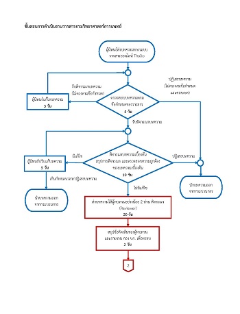
กรมวิทยาศาสตร์การแพทย์. แนวทางการดําเนินงานการขออนุญาตห้องปฏิบัติการรังสีวินิจฉัยทางการแพทย์. [ออนไลน์]. 2565; [สืบค้น 27 พ.ย. 2567]: [92 หน้า]. เข้าถึงได้จาก: URL: https://webapp1.dmsc.moph.go.th/petitionxray/web3/download/XrayTotal.pdf.
ดาวน์โหลด
เผยแพร่แล้ว
รูปแบบการอ้างอิง
ฉบับ
ประเภทบทความ
สัญญาอนุญาต
ลิขสิทธิ์ (c) 2025 วารสารกรมวิทยาศาสตร์การแพทย์

อนุญาตภายใต้เงื่อนไข Creative Commons Attribution-NonCommercial-NoDerivatives 4.0 International License.