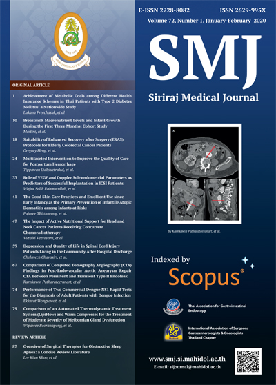
Comparison of Computed Tomography Angiography (CTA) Findings in Post-Endovascular Aortic Aneurysm Repair CTA between Persistent and Transient Type II Endoleak
DOI:
https://doi.org/10.33192/Smj.2020.09Keywords:
Comparison; computed tomography angiography; CTA; post-endovascular aortic aneurysm repair CTA; persistent and transient type II endoleakAbstract
Objective: To compare first post-endovascular aortic aneurysm repair (EVAR) computed tomography angiography (CTA) imaging characteristics between transient and persistent type II endoleaks.
Methods: This retrospective study enrolled patients who underwent EVAR and were diagnosed with type II endoleak from first post-operative CTA during January 2005 to October 2017 at the Department of Radiology, Faculty of Medicine Siriraj Hospital, Mahidol University, Bangkok, Thailand. Aneurysmal sac size, aneurysmal sac growth, and endoleak were recorded among patients whose endoleak disappeared within 6 months (transient group), and among patients whose endoleak persisted for more than 6 months (persistent group).
Results: Eighty-eight patients with a mean age of 75.3±7.3 years were included. Of those, 12 and 76 patients were in the transient group and persistent group, respectively. There were 71 males and 17 females. Univariate analysis showed number of feeding arteries (odds ratio [OR]: 9.9, p=0.012) and presence of inferior mesenteric artery (IMA) as an endoleak source (OR: 4.3, p=0.026) to be found more frequently in the persistent group than in the transient group; however, neither factor survived multivariate analysis. No significant difference between two groups was seen for endoleak diameter, endoleak complexity, or aneurysmal sac enlargement.
Conclusion: The number of feeder arteries and presence of IMA as an endoleak source on first postoperative CTA to be more likely found in patients with persistent type II endoleak. Further prospective study in a larger study population is necessary to identify any existing statistically significant differences and/or associations.
References
2. Lederle FA, Freischlag JA, Kyriakides TC, Matsumura JS, Padberg FTJ, Kohler TR, et al. Long-term comparison of endovascular and open repair of abdominal aortic aneurysm. N Engl J Med 2012;367:1988-97.
3. Jones JE, Atkins MD, Brewster DC, Chung TK, Kwolek CJ, LaMuraglia GM, et al. Persistent type 2 endoleak after endovascular repair of abdominal aortic aneurysm is associated with adverse late outcomes. J Vasc Surg 2007;46:1-8.
4. Lo RC, Buck DB, Herrmann J, Hamdan AD, Wyers M, Patel VI, et al. Risk factors and consequences of persistent type II endoleaks. J Vasc Surg 2016;63:895-901.
5. Christopher J. Abularrage, Robert S. Crawford, Mark F. Conrad, Hang Lee, Christopher J. Kwolek, David C. Brewster, et al. Preoperative Variables Predict Persistent Type 2 Endoleak After Endovascular Aneurysm Repair. J Vasc Surg 2010;52:19-24.
6. Couchet G, Pereira B, Carrieres C, Maumias T, Ribal J-P, Ben Ahmed S, et al. Predictive factors for type II endoleaks after treatment of abdominal aortic aneurysm by conventional endovascular aneurysm repair. Ann Vasc Surg 2015;29:1673-9.
7. Mursalin R, Sakamoto I, Nagayama H, Sueyoshi E, Tanigawa K, Miura T, et al. Imaging-based predictors of persistent type II endoleak after endovascular abdominal aortic aneurysm repair. AJR Am J Roentgenol 2016;206:1335-40.
8. Müller-Wille R, Schötz S, Zeman F, Uller W, Güntner O, Pfister K, et al. CT features of early type II endoleaks after endovascular repair of abdominal aortic aneurysms help predict aneurysm sac enlargement. Radiology 2015;274:906-16.
9. Ward TJ, Cohen S, Patel RS, Kim E, Fischman AM, Nowakowski FS, et al. Anatomic risk factors for type-2 endoleak following EVAR: A retrospective review of preoperative CT angiography in 326 patients. Cardiovasc Intervent Radiol 2014;37:324-8.
10. Maeda T, Ito T, Kurimoto Y, Watanabe T, Kuroda Y, Kawaharada N, et al. Risk factors for a persistent type 2 endoleak after endovascular aneurysm repair. Surg Today 2015;45:1373-7.
11. Timaran CH, Ohki T, Rhee SJ, Veith FJ, Gargiulo NJ, Toriumi H, et al. Predicting aneurysm enlargement in patients with persistent type II endoleaks. J Vasc Surg 2004;39:1157-62.
12. Keedy AW, Yeh BM, Kohr JR, Hiramoto JS, Schneider DB, Breiman RS. Evaluation of potential outcome predictors in type II endoleak: A retrospective study with CT angiography feature analysis. AJR Am J Roentgenol 2011;197:234-40.
13. Dudeck O, Schnapauff D, Herzog L, Löwenthal D, Bulla K, Bulla B, et al. Can early computed tomography angiography after endovascular aortic aneurysm repair predict the need for reintervention in patients with type II endoleak? Cardiovasc Intervent Radiol 2015;38:45-52.
14. Pippin K, Hill J, He J, Johnson P. Outcomes of type II endoleaks after endovascular abdominal aortic aneurysm (AAA) repair: a single-center, retrospective study. Clin Imaging 2016;40:875-9.
Downloads
Published
How to Cite
Issue
Section
License
Authors who publish with this journal agree to the following conditions:
Copyright Transfer
In submitting a manuscript, the authors acknowledge that the work will become the copyrighted property of Siriraj Medical Journal upon publication.
License
Articles are licensed under a Creative Commons Attribution-NonCommercial-NoDerivatives 4.0 International License (CC BY-NC-ND 4.0). This license allows for the sharing of the work for non-commercial purposes with proper attribution to the authors and the journal. However, it does not permit modifications or the creation of derivative works.
Sharing and Access
Authors are encouraged to share their article on their personal or institutional websites and through other non-commercial platforms. Doing so can increase readership and citations.


