
Multimodal Imaging of Unaffected Fellow Eyes in Patients with Polypoidal Choroidal Vasculopathy and Neovascular Age-Related Macular Degeneration
DOI:
https://doi.org/10.33192/Smj.2021.17Keywords:
Multimodal Imaging, Age-related macular degeneration, Choroidal neovascularization, Polypoidal choroidal vasculopathy, Fellow eyes, Retinal imagingAbstract
Objective: To identify retinal abnormalities in the unaffected fellow eyes of patients with unilateral polypoidal choroidal vasculopathy (PCV) and neovascular aged-related macular degeneration (n-AMD).
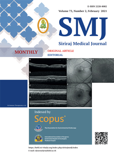
Methods: In this cross-sectional, retrospective case series, the medical records of patients with PCV and n-AMD were reviewed and the baseline patient characteristics recorded. Abnormal findings on spectral-domain optical coherence tomography (SD-OCT) (steep/notched pigment epithelial detachment [PED], double-layer sign, hyporeflective lumen within the PED), fundus autofluorescence (FAF) (ring/patch patterns), and indocyanine green angiography (ICGA) (punctate hyperfluorescence spot [PHS]) were studied.
Results: Seventy-one fellow eyes of patients with PCV and 64 fellow eyes of patients with n-AMD were included. FAF showed abnormalities in 26 (36.6%) and 33 (51.6%) fellow eyes of those with PCV and n-AMD, respectively (p=0.081). SD-OCT detected abnormalities in 25 (35.2%) and 36 (56.3%) fellow eyes of those with PCV and n-AMD, respectively (p=0.014). ICGA detected PHS in 47 (66.2%) and 34 (53.1%) fellow eyes of PCV and n-AMD, respectively (p=0.122).
Conclusion: Multimodal imaging showed abnormalities in most asymptomatic fellow eyes of patients with PCV and n-AMD. Regular and long-term self-monitoring and fundus evaluation are important for these patients. The current findings support the differences in the pathogeneses of PCV and n-AMD.
References
2. Green WR, Enger C. Age-related macular degeneration histopathologic studies. The 1992 Lorenz E. Zimmerman Lecture. Ophthalmology. 1993;100(10):1519-35.
3. Kahn HA, Leibowitz HM, Ganley JP, Kini MM, Colton T, Nickerson RS, et al. The Framingham Eye Study. I. Outline and major prevalence findings. Am J Epidemiol. 1977;106(1):17-32.
4. Cheung CMG, Lee WK, Koizumi H, Dansingani K, Lai TYY, Freund KB. Pachychoroid disease. Eye (Lond). 2019;33(1):14-33.
5. Ferris FL, Davis MD, Clemons TE, Lee LY, Chew EY, Lindblad AS, et al. A simplified severity scale for age-related macular degeneration: AREDS Report No. 18. Arch Ophthalmol. 2005;123(11):1570-4.
6. Chew EY, Clemons TE, Agron E, Sperduto RD, Sangiovanni JP, Davis MD, et al. Ten-year follow-up of age-related macular degeneration in the age-related eye disease study: AREDS report no. 36. JAMA Ophthalmol. 2014;132(3):272-7.
7. Koh A, et al. . EVEREST study: efficacy and safety of verteporfin photodynamic therapy in combination with ranibizumab or alone versus ranibizumab monotherapy in patients with symptomatic macular polypoidal choroidal vasculopathy. Retina 328 (2012): 1453-1464.
8. Argon laser photocoagulation for senile macular degeneration. Results of a randomized clinical trial. Arch Ophthalmol. 1982;100(6):912-8.
9. LI H. YANG M, * JOST B. JONAS, MD,†‡ WEN B. WEI, MD*. OPTICAL COHERENCE TOMOGRAPHIC ENHANCED DEPTH IMAGING OF POLYPOIDAL CHOROIDAL VASCULOPATHY. RETINA 33:1584–1589, 2013.
10. Nandakumar N, Buzney S, Weiter JJ. Lipofuscin and the principles of fundus autofluorescence: a review. Semin Ophthalmol. 2012;27(5-6):197-201.
11. Five-year follow-up of fellow eyes of patients with age-related macular degeneration and unilateral extrafoveal choroidal neovascularization. Macular Photocoagulation Study Group. Archives of ophthalmology. 1993;111(9):1189-99.
12. Age-Related Eye Disease Study Research G. A randomized, placebo-controlled, clinical trial of high-dose supplementation with vitamins C and E, beta carotene, and zinc for age-related macular degeneration and vision loss: AREDS report no. 8. Archives of ophthalmology. 2001;119(10):1417-36.
13. Sandberg MA, Weiner A, Miller S, Gaudio AR. High-risk characteristics of fellow eyes of patients with unilateral neovascular age-related macular degeneration. Ophthalmology. 1998;105(3):441-7.
14. Ueta T, Iriyama A, Francis J, Takahashi H, Adachi T, Obata R, et al. Development of typical age-related macular degeneration and polypoidal choroidal vasculopathy in fellow eyes of Japanese patients with exudative age-related macular degeneration. Am J Ophthalmol. 2008;146(1):96-101.
15. Uyama M, Takahashi K, Ida N, Miyashiro M, Ando A, Takahashi A, et al. The second eye of Japanese patients with unilateral exudative age related macular degeneration. The British journal of ophthalmology. 2000;84(9):1018-23.
16. Schmitz-Valckenberg S, Holz FG, Bird AC, Spaide RF. Fundus autofluorescence imaging: review and perspectives. Retina. 2008;28(3):385-409.
17. Zarbin MA. Age-related macular degeneration: review of pathogenesis. Eur J Ophthalmol. 1998;8(4):199-206.
18. Dansingani KK, Balaratnasingam C, Naysan J, Freund KB. En Face Imaging of Pachychoroid Spectrum Disorders with Swept-Source Optical Coherence Tomography. Retina. 2016;36(3):499-516.
19. Balaratnasingam C, Lee WK, Koizumi H, Dansingani K, Inoue M, Freund KB. Polypoidal Choroidal Vasculopathy: A Distinct Disease or Manifestation of Many? Retina. 2016;36(1):1-8.
20. Lee WK, Baek J, Dansingani KK, Lee JH, Freund KB. Choroidal Morphology in Eyes with Polypoidal Choroidal Vasculopathy and Normal or Subnormal Subfoveal Choroidal Thickness. Retina. 2016;36 Suppl 1:S73-S82.
21. Yamagishi T, Koizumi H, Yamazaki T, Kinoshita S. Fundus autofluorescence in polypoidal choroidal vasculopathy. Ophthalmology. 2012;119(8):1650-7.
22. Tsujikawa A, Ojima Y, Yamashiro K, Ooto S, Tamura H, Nakagawa S, et al. Punctate hyperfluorescent spots associated with central serous chorioretinopathy as seen on indocyanine green angiography. Retina. 2010;30(5):801-9.
23. Kim H, Lee JH, Kwon KY, Byeon SH, Lee SC, Lee CS. Punctate hyperfluorescent spots associated with polypoidal choroidal vasculopathy on indocyanine green angiography. Ophthalmic Surg Lasers Imaging Retina. 2015;46(4):423-7.
24. Park SJ, Kim BH, Park KH, Woo SJ. Punctate hyperfluorescence spot as a common choroidopathy of central serous chorioretinopathy and polypoidal choroidal vasculopathy. Am J Ophthalmol. 2014;158(6):1155-63 e1.
Published
How to Cite
Issue
Section
License
Copyright (c) 2020 Siriraj Medical Journal

This work is licensed under a Creative Commons Attribution-NonCommercial-NoDerivatives 4.0 International License.
Authors who publish with this journal agree to the following conditions:
Copyright Transfer
In submitting a manuscript, the authors acknowledge that the work will become the copyrighted property of Siriraj Medical Journal upon publication.
License
Articles are licensed under a Creative Commons Attribution-NonCommercial-NoDerivatives 4.0 International License (CC BY-NC-ND 4.0). This license allows for the sharing of the work for non-commercial purposes with proper attribution to the authors and the journal. However, it does not permit modifications or the creation of derivative works.
Sharing and Access
Authors are encouraged to share their article on their personal or institutional websites and through other non-commercial platforms. Doing so can increase readership and citations.


