
Change in Corneal Endothelial Cell Density after Baerveldt Shunt Implantation in Glaucoma Patients
DOI:
https://doi.org/10.33192/Smj.2021.33Keywords:
Corneal endothelial cells, Baerveldt shunt implantation, glaucoma, ThailandAbstract
Objective: To evaluate changes in corneal endothelial cells density (EDC) at 1 one, six, and twelve months after Baerveldt shunt implantation.
Materials and Methods: This prospective study included 24 patients who underwent Baerveldt shunt implantation for refractory glaucoma, and who had one full year of post-surgical follow-up. Best corrected visual acuity (BCVA), intraocular pressure (IOP), number of glaucoma medications, central corneal thickness (CCT), corneal endothelial cell density (EDC), and morphology in central, inferior, and superotemporal (stEDC) areas were recorded at baseline, and 1, 6, and 12 months after surgery. Distance between the tip of tube to corneal endothelium (TTC) was measured using optical coherence tomography at one month after surgery.
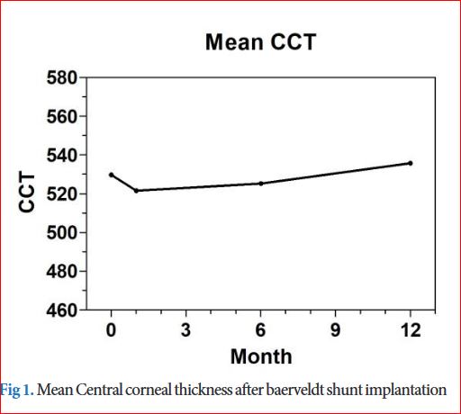
Results: Twenty-four eyes from 24 patients were analyzed. Sixty-two percent were primary open-angle glaucoma, and 73.1% of patients had previous trabeculectomy. Mean BCVA was not significantly changed. The mean IOP at six months (12.2±4.35 mmHg) and at one year (11.1±4.31 mmHg) was significantly lower than baseline (20.1±9.24 mmHg) (p<0.001 and p<0.001 respectively). Median (min, max) number of anti-glaucoma medications significantly decreased from 4 (1, 6) at baseline to 1 (0, 3) and 1 (0, 3) at six months and one year after surgery (p<0.001 and p<0.001, respectively). Mean baseline stEDC was 1,527±644 cells/mm2. From linear mixed model, stEDC showed the most significantly decreasing slope (y=1365.54 – 18.6125t, p=0.014), and CCT showed a significant increase over time (y=533.65 + 1.8853t). Pearson’s correlation coefficient between TTC and stEDC change at one year was not statistically significant (-0.403, p=0.172).
Conclusion: After Baerveldt shunt implantation, EDC loss over time was found in the area closest to where the tube was placed in addition to increasing CCT. Distance from tip of tube to cornea is not the only factor that can cause EDC loss after shunt implantation. Additional study to identify other possible mechanisms is warranted.
Downloads
Published
How to Cite
Issue
Section
License
Authors who publish with this journal agree to the following conditions:
Copyright Transfer
In submitting a manuscript, the authors acknowledge that the work will become the copyrighted property of Siriraj Medical Journal upon publication.
License
Articles are licensed under a Creative Commons Attribution-NonCommercial-NoDerivatives 4.0 International License (CC BY-NC-ND 4.0). This license allows for the sharing of the work for non-commercial purposes with proper attribution to the authors and the journal. However, it does not permit modifications or the creation of derivative works.
Sharing and Access
Authors are encouraged to share their article on their personal or institutional websites and through other non-commercial platforms. Doing so can increase readership and citations.


