
Differentiation Between the Malignant Mesothelioma of Pleura and Pleural Metastasis with Contrast Enhanced CT, Is It Possible?
DOI:
https://doi.org/10.33192/Smj.2021.77Keywords:
Malignant pleural mesothelioma, pleural metastasisAbstract
Objective: To compare CT findings between malignant pleural mesothelioma (MPM) and metastatic pleural
disease (MPD).
Materials and Methods: CT chest images of 157 cases of pathologically-proven malignant pleural disease
(21 MPM, 136 MPD) were retrospectively reviewed by two radiologists who were blinded to the diagnosis. Findings
of interest included pleural effusion, pleural thickening, organ invasion, lymphadenopathy, dominant lung nodule,
pulmonary or extra-thoracic organ metastasis, and asbestos-related disease.
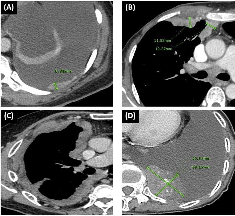
Results: Findings commonly found in MPM compared with MPD are circumferential pleural thickening (52.4%
vs 14.0%, p<0.001), pleural mass (33.3% vs 7.4%, p<0.001), organs invasion (57.1% vs 9.6%, p<0.001), and asbestos
related disease (19% vs 0%, p<0.001).
Conclusions: Circumferential pleural thickening, pleural mass, presence of organ invasion, and CT finding of
asbestos-related pleural disease were the CT findings that raise the possibility of MPM.
References
Price B. Analysis of current trends in United States mesothelioma incidence. Am J Epidemiol 1997; 145:211–218.
Spirtas R, Heineman EF, Bernstein L, et al. Malignant mesothelioma: attributable risk of asbestos exposure. Occup Environ Med 1994; 51:804-811.
Bianchi C, Bianchi T. Malignant mesothelioma: global incidence and relationship with asbestos. Ind Health. 2007 Jun; 45(3):379- 87
Ioannis Psallidas, Ioannis Kalomenidis, Jose M. Porcel, Bruce
W. Robinson, Georgios T. Stathopoulos. Malignant pleural
effusion: from bench to bedside. European Respiratory Review Jun 2016, 25 (140) 189-198.
Johnston WW. The malignant pleural effusion. A review of cytopathologic diagnoses of 584 specimens from 472 consecutive patients. Cancer 1985; 56:905-910.
Rusch V.W, Kari Chansky, MS,b Hedy L. Kindler, MD,c Anna K. Nowak, M.B.B.S., PhD, et al. THE IASLC Mesothelioma Staging Project: Proposals for the M Descriptos and for Revision of the TNM Stage Groupings in the Forthcoming (Eighth) Edition of the TNM Classification for Mesothelioma. Journal of Thoracic Oncology 2016, Vol 11, No 12, 2112-2119.
Burrows CM, Mathews WC, Colt HG. Predicting survival in patients with recurrent symptomatic malignant pleural effusions: an assessment of the prognostic values of physiologic, morphologic, and quality of life measures of extent of disease. Chest 2000;117: 73-8.
Bielsa S, Salud A, Martinez M, et al. Prognostic significance of pleural fluid data in patients with malignant effusion. Eur J Intern Med 2008;19: 334-9.
Arnaud Scherpereel, Isabelle Opitz, Thierry Berghmans, Ioannis Psallidas, Markus Glatzer, et al. ERS/ESTS/EACTS/ESTRO guidelines for the management of malignant pleural mesothelioma. European Respiratory Journal Jan 2020, 1900953
Scherpereel A, Astoul P, Baas P, et al. Guidelines of the European Respiratory Society and the European Society of Thoracic Surgeons for the management of malignant pleural mesothelioma. Eur Respir J 2010;35: 479-95.
Woolhouse I, Bishop L, Darlison L, De Fonseka D, Edey A, Edwards J, Faivre-Finn C, Fennell DA, Holmes S, Kerr KM, Nakas A. British Thoracic Society Guideline for the investigation and management of malignant pleural mesothelioma. Thorax. 2018 Mar 1;73(Suppl 1): i1-30.
Seely JM, Nguyen ET, Churg AM, et al. Malignant pleural mesothelioma: computed tomography and correlation with histology. Eur J Radiol 2009;70(3):485-91.
Metintas M, Ucgun I, Elbek O, et al. Computed tomography features in malignant pleural mesothelioma and other commonly seen pleural diseases. Eur J Radiol 2002;41(1):1-9.
Leung AN, Muller NL, Miller RR. CT in differential diagnosis of diffuse pleural disease. AJR Am J Roentgenol 1990;154(3):487-92.
Yilmaz U, Polat G, Sahin N, et al. CT in differential diagnosis of benign and malignant pleural disease. Monaldi Arch Chest Dis 2005;63: 17–22
Senyigit A, Bayram H. Malignant pleural mesothelioma caused by environmental exposure to asbestos in the Southeast of Turkey: CT findings in 117 patients. Respiration. 2000;67(6):615–22
Yoon K et al. Multidetector CT Findings and Differential Diagnoses of Malignant Pleural Mesothelioma and Metastatic Pleural Diseases in Korea, Korean J Radiol 2016;17(4):545-553
Mehrdad B K. Malignant Mesothelioma Versus Metastatic Carcinoma of the Pleura: A CT Challenge, Iran J Radiol. 2016 January; 13(1): e10949
Sheila D. Davis, MD. CT Evaluation for Pulmonary Metastases in Patients with Extrathoracic Malignancy. Radiology 1991;180: 1-12 602 Volume 73, No.9: 2021 Siriraj Medical Journal https://he02.tci- thaijo.org/index.php/sirirajmedj/index
Gross BH, Glazer GM, Bookstein FL. Multiple pulmonary nodules detected by computed tomography: diagnostic implications. J Comput Assist Tomogr 1985; 9:880-885.
Dorfman RE, Alpern MB, Gross BH, Sandler MA. Upper abdominal lymph nodes: criteria for normal size determined with CT. Radiology 1991; 180:319 –322
Ashley R. Cahoon, MD, Benjamin D. Smith, MD, Wei T. Yang, MBBS. Internal Thoracic Lymphadenopathy in Breast Cancer, RadioGraphics 2017; 37:1024–1036
Tamer Dogan O, Salk I, Tas F, Epozturk K, Gumus C, AkkurtI, et al. Thoracic Computed Tomography Findings in Malignant Mesothelioma. Iran J Radiol 2012; 9 (4): 209-11.
Peacock C, Copley SJ, Hansell DM. Asbestosrelated benign pleural disease. Clin Radiol 2000; 55:422–432.
Henschke CI, Yankelevitz DF, Davis SD. Pleural diseases: multimodality imaging and clinical management. Cunr Probl Diagn Radiol 1991; 20:159-179.
Mongardon N, Pinton-Gonnet C, Szekely B, et al. Assessment of chronic pain after thoracotomy: a 1-year prevalence study. Clin J Pain 2011; 27: 677–681.
Coppage L, Shaw C. Curtis AM. Metastatic disease to the chest in patients with extrathoracic malignancies. J Thorac Imaging. 1987; 2: 24–37
Johkoh T, Ikezoe J, Tomiyama N et al. CT findings in lymphangitic carcinomatosis of the lung: correlation with histologic findings and pulmonary function tests. AJR 1992; 158: 1217–22.
Rahman NM, Wang NS. Anatomy of the pleura. In: Light RW, Lee YCG, eds. Textbook of pleural diseases. 2nd ed. Boca Raton, Fla: CRC, 2008; 13–25.
Negrini D, Moriondo A. Pleural function and lymphatics. Acta Physiol (Oxf) 2013;207(2):244–259.
Abdel Rahman AR, Gaafar RM, Baki HA, El Hosieny HM, Aboulkasem F, Farahat EG, et al. Prevalence and pattern of lymph node metastasis in malignant pleural mesothelioma. Ann Thorac Surg 2008;86:391-395
Downloads
Published
How to Cite
Issue
Section
License
Authors who publish with this journal agree to the following conditions:
Copyright Transfer
In submitting a manuscript, the authors acknowledge that the work will become the copyrighted property of Siriraj Medical Journal upon publication.
License
Articles are licensed under a Creative Commons Attribution-NonCommercial-NoDerivatives 4.0 International License (CC BY-NC-ND 4.0). This license allows for the sharing of the work for non-commercial purposes with proper attribution to the authors and the journal. However, it does not permit modifications or the creation of derivative works.
Sharing and Access
Authors are encouraged to share their article on their personal or institutional websites and through other non-commercial platforms. Doing so can increase readership and citations.


