
Temporal Bone Landmarks of the Transverse-sigmoid Sinus Junction: An Anatomical Study in Dried Human Skulls
DOI:
https://doi.org/10.33192/Smj.2021.95Keywords:
Relationship; transverse-sigmoid sinus junction, squamosal suture; parietomastoid suture, supramastoid crest, temporal craniotomy, middle cranial fossaAbstract
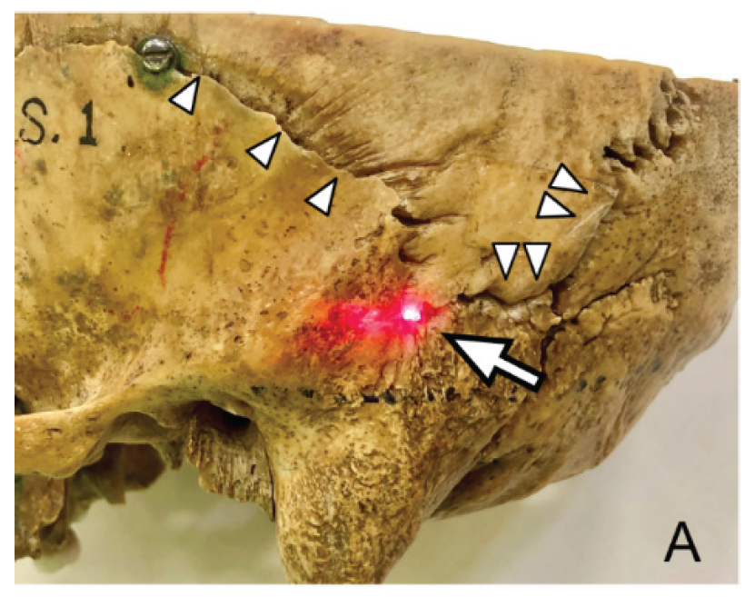
Objective: To investigate the accuracy in localization of the anterosuperior margin of TSSJ by using the intersection point between the squamosal and parietomastoid sutures (A point) and the intersection of the squamosal suture and supramastoid crest (B point) as bony landmarks.
Materials and Methods: The A and B points were marked on the inner surface of a skull by using the transillumination technique. The anatomical relationship between the projected A point, B point, and groove of TSSJ was investigated in 60 dried Thai human skulls (120 sides).
Results: Of the 120 sides, the projected A points were located exactly on the anterosuperior margin of the TSSJ in 38 (31.7%) instances and adjacent (above and below) the anterosuperior margin in 82 (68.3%) cases. Of the 118 sides with identifiable supramastoid crests, the projected B points were located precisely on the anterosuperior margin of TSSJ in 60 (50.8%) cases and above the anterosuperior margin of the TSSJ in 57 (48.3%) cases. Hence, the projected B point was a more reliable bony landmark for localizing the anterosuperior margin of the TSSJ when compared with the projected A point (p = 0.003, OR 2.2, and 95% CI =1.3-3.8).
Conclusion: The B point is a more reliable temporal bone landmark for localization of the TSSJ than the A point. In temporal craniotomy, an initial burr hole at the B point is relatively safe and carries a very low risk of inadvertent venous sinus injury.
References
2. Raza SM, Quinones-Hinojosa A. The extended retrosigmoid approach for neoplastic lesions in the posterior fossa: technique modification. Neurosurg Rev. 2011;34(1):123-9.
3. Dogan I, Ozgural O, Eroglu U, Al-Beyati ESM, Kilinc CM, Comert A, et al. Preoperative exposure of sigmoid sinus trajectory in posterolateral cranial base approaches using a new landmark through a neurosurgical perspective. J Craniofac Surg. 2018;29(1):220-5.
4. Ugur HC, Dogan I, Kahilogullari G, Al-Beyati ES, Ozdemir M, Kayaci S, et al. New practical landmarks to determine sigmoid sinus free zones for suboccipital approaches: an anatomical study. J Craniofac Surg. 2013;24(5):1815-8.
5. Ucerler H, Govsa F. Asterion as a surgical landmark for lateral cranial base approaches. J Craniomaxillofac Surg. 2006;34(7):415-20.
6. Bozbuga M, Boran BO, Sahinoglu K. Surface anatomy of the posterolateral cranium regarding the localization of the initial burr–hole for a retrosigmoid approach. Neurosurgical Review. 2006;29(1):61-3.
7. Day JD, Jordi XK, Manfred T, Takanori F. Surface and superficial surgical anatomy of the posterolateral cranial base: significance for surgical planning and approach. Neurosurgery. 1996;38(6):1079-84.
8. Li RC, Liu JF, Li K, Qi L, Yan SY, Wang MD, et al. Localization of anterosuperior point of transverse-sigmoid sinus junction using a reference coordinate system on lateral skull surface. Chin Med J (Engl). 2016;129(15):1845-9.
9. Goto T, Ishibashi K, Morisako H, Nagata T, Kunihiro N, Ikeda H, et al. Simple and safe exposure of the sigmoid sinus with presigmoid approaches. Neurosurg Rev. 2013;36:477‑82.
10. Sheng B, Lv F, Xiao Z, Ouyang Y, Lv F, Deng J, et al. Anatomical relationship between cranial surface landmarks and venous sinus in posterior cranial fossa using CT angiography. Surg Radiol Anat. 2012;34(8):701-8.
11. Blumenfeld J. Racial identification in the skull and teeth. The University of Western Ontario Journal of Anthropology 2000;8(1).
12. Low WK, Fenton JE, Fagan PA, Gibson WP. Racial considerations in acoustic neuroma removal with hearing preservation via the retrosigmoid approach. Acta Otolaryngol. 1995;115(6):783-6.
13. Duangthongpon P, Thanapaisal C, Kitkhuandee A, Chaiciwamongkol K, Morthong V. Supramastoid crest, safety landmark for craniotomy? J Med Assoc Thai. 2013;96(4):S138-41.
14. Tomaszewska A, Bisiecka A, Pawelec Ł. Asterion localization-variability of the location for surgical and anthropological relevance. Homo. 2020;70(4):325-33.
15. Day JD, Tschabitscher M. Anatomic position of the asterion. Neurosurgery. 1998;42(1):198-9.
16. Sripairojkul B, Adultrakoon A. Anatomical position of the asterion and its underlying structure. J Med AssocThai. 2000;83(9):1112-5.
17. Bellary SS, Steinberg A, Mirzayan N, Shirak M, Tubbs RS, Cohen‐Gadol AA, Loukas M. Wormian bones: a review. Clin Anat. 2013;26(8):922-7.
18. Gharehdaghi J, Jafari-Marandi H, Faress F, Zeinali M, Safari H. Morphology of asterion and its proximity to deep vein sinuses in Iranian adult skull. Br J Neurosurg. 2020;34(1):55-58.
19. Sudha R, Sridevi C, Ezhilarasi M. Anatomical variations in the formation of pterion and asterion in South Indian population. Int J Cur Res Rev. 2013;5(09):92-101.
20. Ghosh SK, Biswas S, Sharma S, Chakraborty S. An anatomical study of wormian bones from the eastern part of India: is genetic influence a primary determinant of their morphogenesis?. Anat Sci Int. 2017;92(3):373-82.
21. Johnson DR, O'higgins P, Moore WJ, McAndrew TJ. Determination of race and sex of the human skull by discriminant function analysis of linear and angular dimensions. Forensic Sci Int. 1989;41(1-2):41-53.
22. Dekaban AS. Tables of cranial and orbital measurements, cranial volume, and derived indexes in males and females from 7 days to 20 years of age. Ann Neurol. 1977;2(6):485-91.
23. Albert AM, Ricanek Jr K, Patterson E. A review of the literature on the aging adult skull and face: Implications for forensic science research and applications. Forensic Sci Int. 2007;172(1):1-9.
24. Nikita E. Αge‐associated variation and sexual dimorphism in adult cranial morphology: Implications in anthropological studies. Int J Osteoarchaeol. 2014;24(5):557-69.
25. Gapert R, Black S, Last J. Test of age-related variation in the craniometry of the adult human foramen magnum region: implications for sex determination methods. Forensic Sci Med Pathol. 2013;9(4):478-88.
Published
How to Cite
Issue
Section
License
Authors who publish with this journal agree to the following conditions:
Copyright Transfer
In submitting a manuscript, the authors acknowledge that the work will become the copyrighted property of Siriraj Medical Journal upon publication.
License
Articles are licensed under a Creative Commons Attribution-NonCommercial-NoDerivatives 4.0 International License (CC BY-NC-ND 4.0). This license allows for the sharing of the work for non-commercial purposes with proper attribution to the authors and the journal. However, it does not permit modifications or the creation of derivative works.
Sharing and Access
Authors are encouraged to share their article on their personal or institutional websites and through other non-commercial platforms. Doing so can increase readership and citations.


