
Age-Related Changes in Signal Intensity Ratio of Normal Clivus Bone Marrow on Magnetic Resonance Imaging
DOI:
https://doi.org/10.33192/Smj.2022.46Keywords:
Clivus bone marrow, pons, signal intensity ratio, magnetic resonance imagingAbstract
Objective: To evaluate the association between the signal intensity ratio of clivus bone displayed on magnetic resonance (MR) imaging and ages.
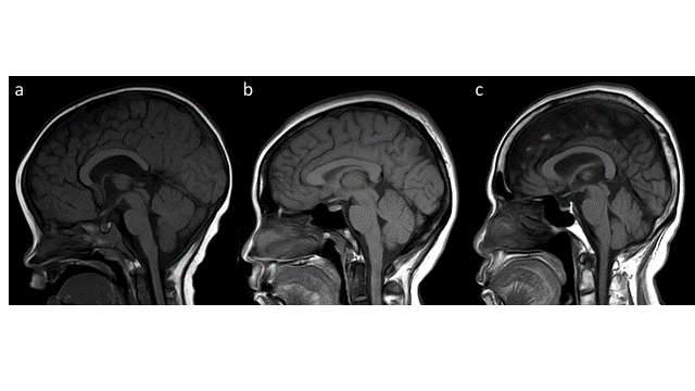
Materials and Methods: A retrospective cohort study of 268 patients underwent brain MR imaging during January 2015 to October 2019. We qualitatively and quantitatively assessed bone marrow signal intensity of clivus bone that were performed on T1-weighted sagittal images. In qualitative assessment, the signal intensities of clivus were visually graded from Grade I to III according to the proportion of low and high signal intensity areas occupying the clival marrow region. In quantitative assessment, we evaluated the association between the signal intensity ratio of clivus to pons and age categorized by decades in multivariable Gaussian regression analysis.
Results: Of 268 patients, the ratio of males to females is 1:1. Grade I clivus was found about 35% of the age 1-9 years, whereas Grade 3 clivus was more frequent (more than 13%) in the ages over 30 years. There were statistically different in the mean values of clivus/CSF and clivus/pons signal intensity ratios by grades. The mean values of clivus/CSF and clivus/pons signal intensity ratios were increased by ages in both sexes, but slightly higher in males. In regression analysis after adjustment for sex, the differences in mean values of clivus/pons signal intensity ratios were larger by increasing age, using the age 1-9 as a reference group.
Conclusion: The present study confirms that signal intensity ratios of clivus to pons on T1-weighted sagittal MR images is increased with ages.
References
Olcu E, Arslan M, Sabanciogullari V, Salk I, Marrow B. Magnetic Resonance Imaging of the Clivus and its Age-Related Changes in the Bone Marrow. Iran J Radiol. 2011;8(4):224-9.
Bayramoğlu A, Aydingöz Ü, Hayran M, Öztürk H, Cumhur M. Comparison of qualitative and quantitative analyses of agerelated changes in clivus bone marrow on MR imaging. Clin Anat. 2003;16(4):304-8.
Kimura F, Kim KW, Friedman H, Russell EJ, Breit R. MR imaging of the normal and abnormal clivus. Am J Roentgenol. 1990;155(6):1285-91.
Loevner LA, Tobey JD, Yousem DM, Sonners AI, Hsu WC. MR imaging characteristics of cranial bone marrow in adult patients with underlying systemic disorders compared with healthy control subjects. Am J Neuroradiol. 2002;23(2):248-54.
Churojana A, Lakkhanawat S, Chailerd O, Boonchai T, Cognard C. Cranial dural arteriovenous fistulas: Can noninvasive imaging predict angiographic findings? Siriraj Med J. 2018;70(4):289-97.
Piyapittayanan S, Segsarnviriya C, Ngamsombat C, Cheunsuchon P, Charnchaowanish P, Sc B, et al. Comparison between Dynamic Contrast-Enhanced MRI and Dynamic Susceptibility Contrast MRI in Glioma Grading. Siriraj Med J. 2017;69(6):369-76.
Małkiewicz A, Dziedzic M. Bone marrow reconversion - Imaging of physiological changes in bone marrow. Polish J Radiol. 2012;77(4):45-50.
Chan BY, Gill KG, Rebsamen SL, Nguyen JC. MR imaging of pediatric bone marrow. Radiographics. 2016;36(6):1911-30.
Simonson TM, Kao SCS. Normal childhood developmental patterns in skull bone marrow by MR imaging. Pediatr Radiol. 1992;22(8):556-9.
Roberts CC, Morrison WB, Bancroft LW, Chew FS. Bone marrow changes on MRI: Self-assessment module. Am J Roentgenol. 2009;193(3 Suppl):5-9.
Tinnut S, Galassi W, Oilmungmool N, Chattrapiban T. Agreement on grading of normal clivus using magnetic resonance imaging among radiologists. Eur J Radiol Open [Internet]. 2022;9:100395. Available from: https://doi.org/10.1016/j.ejro.2022.100395
Taccone A, Oddone M, Occhi M, Dell’Acqua A, Ciccone MA. MRI “road-map” of normal age-related bone marrow - I. Cranial bone and spine. Pediatr Radiol. 1995;25(8):588-95.
Vande Berg BC, Malghem J, Lecouvet FE, Maldague B. Magnetic resonance imaging of normal bone marrow. Eur Radiol. 1998;8(8):1327-34.
Vogler JB, Murphy WA. Bone marrow imaging. Radiology. 1988;168(3):679-93.
Laor T, Jaramillo D. MR imaging insights into skeletal maturation: What is normal? Radiology. 2009;250(1):28–38.
Oyar O, Govsa F, Sener RN K. Assessment of normal clivus related to age with magnetic resonance imaging. Surg Radiol Anat. 1996;18:47-49.
Okada Y, Aoki S, Barkovich AJ, Nichimura K, Norman D, Kjos BO BR. Cranial bone marrow in children : Assessment of normal development with MR imaging. Radiology. 1989;176:161-4.
Published
How to Cite
Issue
Section
License
Copyright (c) 2022 Siriraj Medical Journal

This work is licensed under a Creative Commons Attribution-NonCommercial-NoDerivatives 4.0 International License.
Authors who publish with this journal agree to the following conditions:
Copyright Transfer
In submitting a manuscript, the authors acknowledge that the work will become the copyrighted property of Siriraj Medical Journal upon publication.
License
Articles are licensed under a Creative Commons Attribution-NonCommercial-NoDerivatives 4.0 International License (CC BY-NC-ND 4.0). This license allows for the sharing of the work for non-commercial purposes with proper attribution to the authors and the journal. However, it does not permit modifications or the creation of derivative works.
Sharing and Access
Authors are encouraged to share their article on their personal or institutional websites and through other non-commercial platforms. Doing so can increase readership and citations.


