
Deep Peroneal Nerve: Orientation and Branching at the Ankle and Proximal Part of the Foot
DOI:
https://doi.org/10.33192/Smj.2022.53Keywords:
Deep peroneal nerve, inferior extensor retinaculum, anterior tarsal tunnel, dorsalis pedis artery, hallucis longus tendonAbstract
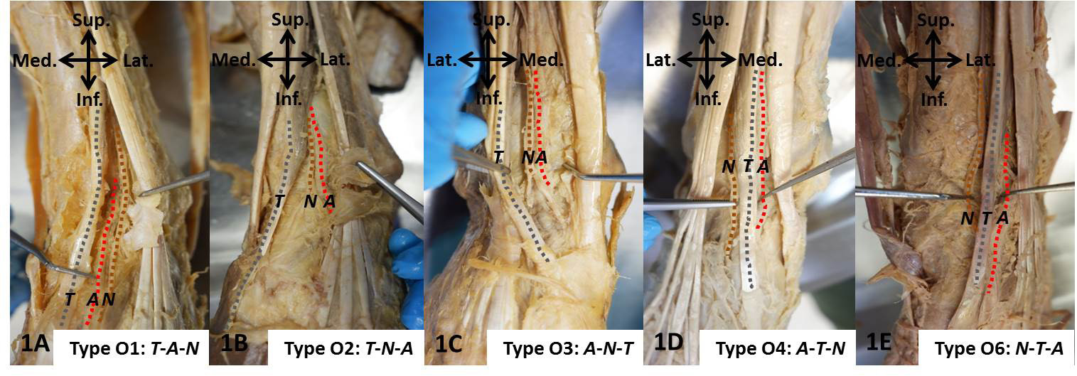
Objective: This study investigated the frequency and types of 1) orientation of the deep peroneal nerve (DPN) and its branches relative to the dorsalis pedis artery (DPA) and the extensor hallucis longus tendon (EHLT) and 2) branching site and pattern of DPN at the distal area of leg and the proximal zone of the foot.
Materials and Methods: One-hundred and sixty specimens from the lower extremities of 80 formalin-embalmed cadavers were investigated for anatomical position, orientation and the branching pattern of DPN by manual dissection, starting from the anterior side of lower extremity just proximal to ankle joint down to the area distal to inferior extensor retinaculum.
Results: The most prevalent medial-to-lateral orientation of structures in the area anterior to ankle joints was the EHLT/DPA/DPN. Comparing DPA with the branching of DPN in the areas inside anterior tarsal tunnel (ATT) and distal to ATT, the most common type was an orientation of DPA that was lateral to both the DPN main trunk and its medial terminal branch. Regarding branching sites and patterns of DPN in the intermalleolar and ATT areas, nearly half of the studied specimens had DPN bifurcation at the intermalleolar level and more than half of the bifurcations were inside the ATT.
Conclusion: This study establishes novel data regarding type variation and prevalence of DPN in areas of ankle and proximal part of foot in the Thai population which could be helpful in clinical practice.
References
Standring S. Gray’s anatomy: the anatomical basis of clinical practice. 41st ed. Philadelphia: Elsevier; 2016.
Lawrence SJ, Botte MJ. The deep peroneal nerve in the foot and ankle: an anatomic study. Foot Ankle Int. 1995;16(11):724-8.
Ikiz ZAA, Ucerler H, Uygur M. The clinical importance of the relationship between the deep peroneal nerve and the dorsalis pedis artery on the dorsum of the foot. Plast Reconstr Surg. 2007;120(3):690-6.
Ranade AV, Rajanigandha V, Rai R, Ebenezer DA. Relationship between the deep peroneal nerve and dorsalis pedis artery in the foot: a cadaveric study. Clin Anat. 2008;21(7):705-12.
Rab M, Ebmer J, Dellon AL. Innervation of the sinus tarsi and implications for treating anterolateral ankle pain. Ann Plast Surg. 2001;47(5):500-4.
Aktan Ikiz ZA, Ucerler H, Uygur M. Dimensions of the anterior tarsal tunnel and features of the deep peroneal nerve in relation to clinical application. Surg Radiol Anat. 2007;29(7):527-30.
Chitra R. The relationship between the deep fibular nerve and the dorsalis pedis artery and its surgical importance. Indian J Plast Surg. 2009;42(1):18-21.
Geller M, Barbato D. Nervus peronaeus profundus. Terminal branches and their variations. Hospital (Rio J). 1970;77(2):679-98.
Andresen BL, Wertsch JJ, Stewart WA. Anterior tarsal tunnel syndrome. Arch Phys Med Rehabil. 1992;73(11):1112-7.
Reed SC, Wright CS. Compression of the deep branch of the peroneal nerve by the extensor hallucis brevis muscle: a variation of the anterior tarsal tunnel syndrome. Can J Surg. 1995;38(6):545-6.
Dellon AL. Deep peroneal nerve entrapment on the dorsum of the foot. Foot Ankle. 1990;11(2):73-80.
Lambert EH. The accessory deep peroneal nerve. A common variation in innervation of extensor digitorum brevis. Neurology. 1969;19(12):1169-76.
Murad H, Neal P, Katirji B. Total innervation of the extensor digitorum brevis by the accessory deep peroneal nerve. Eur J Neurol. 1999;6(3):371-3.
Kudoh H, Sakai T, Horiguchi M. The consistent presence of the human accessory deep peroneal nerve. J Anat. 1999;194(Pt 1):101-8.
Prakash, Bhardwaj AK, Singh DK, Rajini T, Jayanthi V, Singh G. Anatomic variations of superficial peroneal nerve: clinical implications of a cadaver study. Ital J Anat Embryol. 2010;115(3):223-8.
Tomaszewski KA, Roy J, Vikse J, Pekala PA, Kopacz P, Henry BM. Prevalence of the accessory deep peroneal nerve: A cadaveric study and meta-analysis. Clin Neurol Neurosurg. 2016;144:105-11.
Sinanovic O, Zukic S, Muftic M, Tinjic N. Prevalence of Accessory Deep Peroneal Nerve in Sample of Bosnia and Herzegovina Subjects: an Electrophysiological Study. Acta Inform Med. 2021;29(3):193-6.
Crutchfield CA, Gutmann L. Hereditary aspects of accessory deep peroneal nerve. J Neurol Neurosurg Psychiatry. 1973;36(6):989-90.
George A, Alex L, George A. Variations in the origin of dorsalis pedis artery. Indian Journal of Clinical Anatomy and Physiology. 2021;7(4):354-62.
Parikh S, Dawe E, Lee C, Whitehead-Clarke T, Smith C, Bendall S. A cadaveric study showing the anatomical variations in the branches of the dorsalis pedis artery at the level of the ankle joint and its clinical implication in ankle arthroscopy. Ann R Coll Surg Engl. 2017;99(4):286-8.
Rimchala C, Chuckpaiwong B. Relationship of the dorsalis pedis artery to the tarsal navicular. J Foot Ankle Surg. 2015;54(1):66-8.
Ntuli S, Nalla S, Kiter A. Anatomical variation of the Dorsalis pedis artery in a South African population - A Cadaveric Study. Foot (Edinb). 2018;35:16-27.
Hemamalini, Manjunatha HN. Variations in the origin, course and branching pattern of dorsalis pedis artery with clinical significance. Sci Rep. 2021;11(1):1448.
Chompoopong S, Apinhasmit W, Sangiampong A, Amornmettajit N, Charoenwat B, Rattanathamsakul N, et al. Anatomical considerations of the deep peroneal nerve for biopsy of the proximal fibula in Thais. Clin Anat. 2009;22(2):256-60.
Johnston S, Kraus J, Tutton S, Symanski J. Ultrasound-guided diagnostic deep peroneal nerve blocks prior to potential neurectomy: a retrospective review. Skeletal Radiol. 2020;49(8):1313-21.
Fletcher T, Orgill BD, Barth B. Deep Peroneal Nerve Block. StatPearls. Treasure Island (FL)2022.
Lo YL, Leoh TH, Dan YF, Tan YE, Nurjannah S, Fook-Chong S. An electrophysiological study of the deep peroneal sensory nerve. Eur Neurol. 2003;50(4):244-7.
Kim KH, Kim DH, Yun HS, Park BK, Jang JE. Optimal stimulation site for deep peroneal motor nerve conduction study around the ankle: cadaveric study. Ann Rehabil Med. 2012;36(2):182-6.
Koshima I, Nanba Y, Tsutsui T, Takahashi Y. Deep peroneal nerve transfer for established plantar sensory loss. J Reconstr Microsurg. 2003;19(7):451-4.
Akaranuchat N. Lower Extremity Reconstruction with Vascularized Free-Tissue Transfer: 20 Years of Experience in the Faculty of Medicine Siriraj Hospital, Mahidol University, Bangkok, Thailand. Siriraj Med J. 2021;73(7):462-70.
Lui TH. Extensor tendons and deep peroneal nerve adhesion: Treated by complete anterior ankle arthroscopic capsulotomy. Foot Ankle Surg. 2012;18(1):e1-3.
Becciolini M, Pivec C, Riegler G. Ultrasound Imaging of the Deep Peroneal Nerve. J Ultrasound Med. 2021;40(4):821-38.
Sakci Z, Aydin F, Tuncer K, Ogul H. Demonstration With Three-Dimensional Volumetric Magnetic Resonance Sequences of Deep Peroneal Nerve Compression on Os Intermetatarseum Syndrome. Am J Phys Med Rehabil. 2021;100(8):e116-e7.
Meyerkort DJ, Gurel R, Maor D, Calder JDF. Deep Peroneal Nerve Injury Following Hardware Removal
Published
How to Cite
Issue
Section
License

This work is licensed under a Creative Commons Attribution-NonCommercial-NoDerivatives 4.0 International License.
Authors who publish with this journal agree to the following conditions:
Copyright Transfer
In submitting a manuscript, the authors acknowledge that the work will become the copyrighted property of Siriraj Medical Journal upon publication.
License
Articles are licensed under a Creative Commons Attribution-NonCommercial-NoDerivatives 4.0 International License (CC BY-NC-ND 4.0). This license allows for the sharing of the work for non-commercial purposes with proper attribution to the authors and the journal. However, it does not permit modifications or the creation of derivative works.
Sharing and Access
Authors are encouraged to share their article on their personal or institutional websites and through other non-commercial platforms. Doing so can increase readership and citations.


