
Human Maxillary Sinus Development, Pneumatization, Anatomy, Blood Supply, Innervation and Functional Theories: An Update Review
DOI:
https://doi.org/10.33192/Smj.2022.56Keywords:
Human maxillary sinus development, maxillary sinus anatomy, maxillary sinus morphology, theories of paranasal sinus functions, mucous membraneAbstract
The maxillary sinus is one of four pair’s air-filled spaces that surround the nasal cavity. The majority of previously published researches considering the maxillary sinus commonly concentrated on its pathological conditions and the recent surgical or medical management procedures. However, to understand the diseases of this sinus in a better manner, it is essential to have some basic medical details. Therefore, the present review is an attempt to focus light on this sinus by doing a cosmopolitan update on its development and anatomy, besides the functional theories by searching through many well-known scientific databases including Medline, Scopus, EMBASE, PubMed Central (PMC), PubMed, Cochrane Library, and Web of Science.
In the current review, the author tried to approach, recognize and explain the fundamental data that may provide better ideas to imagine the pathological problems related to the maxillary sinus. In addition, the author did not conduct a new study on human nor animal subjects because the previously implemented articles were the chief and the only source for this review.
In conclusion, the functions and physiology of the maxillary sinus are the subjects that reflect the anatomical complexity mentioned in the most recent articles may probably show that all its functions could be a part of a large and more complicated image than that apparent nowadays.
References
Harle F. The history of maxillary sinus surgery from Leonardo da Vinci until today. Bull Hist Dent. 1992;40(2):79-84.
Kaluskar SK. Evolution of Rhinology. Indian J Otolaryngol Head Neck Surg. 2008; 60: 101-5.
Tange RA. Some historical aspects of the surgical treatment of the infected maxillary sinus. Rhinology. 1991;29(2):155-62.
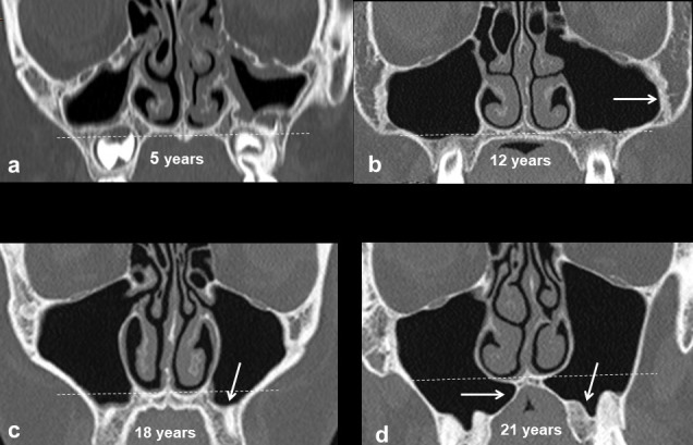
Lawson W, Patel ZM, Lin FY. The development and pathologic processes that influence maxillary sinus pneumatization. Anat Record. 2008;291(11):1554-63.
Bhushan B, Rychlik K, Schroeder JW. Development of the maxillary sinus in infants and children. Int J Pediatr Otorhinolaryngol. 2019;91:146-51.
Przystańska A, Kulczyk T, Rewekant A, Sroka A, Jończyk-Potoczna K, Gawriołek K, et al. The Association between Maxillary Sinus Dimensions and Midface Parameters during Human Postnatal Growth. Biomed Res Int. 2018;18:6391465.
Mohammad SA, Abdalla MA, Mahdi AJJ. Orbitometry of orbital opening and orbital cavity in neonate compared adult. Tikrit Med J. 2011;17:210-6.
Standring S. Gray’s anatomy: the anatomical basis of clinical practice. 42nd ed. London: Elsevier Health Sciences; 2020.
Abdalla MA. Pneumatization patterns of human sphenoid sinus associated with the internal carotid artery and optic nerve by CT scan. Ro J Neurol. 2020;19(4):244-51.
Whyte A, Boeddinghaus R. The maxillary sinus: physiology, development and imaging anatomy. Dentomaxillofac Radiol. 2019;48(8):20190205.
Alqahtani S, Alsheraimi A, Alshareef A, Alsaban R, Alqahtani A, Almgran M, et al. Maxillary Sinus Pneumatization Following Extractions in Riyadh, Saudi Arabia: A Cross-sectional Study. Cureus. 2020;12(1):e6611.
Levi I, Halperin-Sternfeld M, Horwitz J, Zigdon-Giladi H, Machtei EE. Dimensional changes of the maxillary sinus following tooth extraction in the posterior maxilla with and without socket preservation. Clin Implant Dent Relat Res. 2017;19:952-8.
Abdalla MA. Age differences of human sphenoid sinus dimensions: A comparative study by gross anatomical dissection and CT scan imaging. Medicina Moderna. 2021;28(3):321-328.
Neychev D, Kanazirska P, Simitchiev K, Yordanov G. CBCT images: an important tool in the analysis of anatomical variations of maxillary sinus related to Underwood septa features. Biotechnol Biotechnol Equip. 2017;31(6):1210-5.
Fernandes CL. Forensic ethnic identification of crania: the role of the maxillary sinus- a new approach. Am J Forensic Med Pathol. 2004;25(4):302-13.
Abdalla MA, Mahdi AJJ. Maxillary Sinus Measurements in Different Age Groups of Human Cadavers. Tikrit J Dent Sci. 2013;1:107-12.
Maspero C, Farronato M, Bellincioni F, Annibale A, Machetti J, Abate A, et al. Three-Dimensional Evaluation of Maxillary Sinus Changes in Growing Subjects: A Retrospective Cross-Sectional Study. Materials. 2020;13:1007.
Aksoy U, Orhan K. Association between odontogenic conditions and maxillary sinus mucosal thickening: a retrospective CBCT study. Clin Oral Investig. 2019; 23(1):123-31.
Reid L, Meyrick B, Antony VB, Chang LY, Crapo JD, Reynolds HY. The mysterious pulmonary brush cell: a cell in search of a function. Am J Respir Crit Care Med. 2005;172(1):136-9.
Hadar T, Yaniv E, Shvili Y, Koren R, Shvero J. Histopathological changes of the nasal mucosa induced by smoking. Inhal Toxicol. 2009;21(13):1119-22.
Sato K, Chitose SI, Sato K, Sato F, Ono T, Umeno H. Histopathology of maxillary sinus mucosa with odontogenic maxillary sinusitis. Laryngoscope Investig Otolaryngol. 2020;5(2):205-9.
Scherzad A, Hagen R, Hackenberg S. Current Understanding of Nasal Epithelial Cell Mis-Differentiation. J Inflamm Res. 2019;12:309-17.
Harkema JR, Carey SA, Wagner JG, Dintzis SM, Liggitt D. Nose, Sinus, Pharynx, and Larynx. Comp Anat Histol. 2018;1:89-114.
Hung K, Montalvao C, Yeung AW, Li G, Bornstein MM. Frequency, location, and morphology of accessory maxillary sinus ostia: a retrospective study using cone beam computed tomography (CBCT). Surg Radiol Anat. 2020;42(2):219-28.
Abdalla MA. Maxillary Sinus Dimensions of Different Human Age Groups by CT Scan Imaging. Medicina Moderna. 2021;28(2):235-41.
Papadopoulou AM, Chrysikos D, Samolis A, Tsakotos G, Troupis T. Anatomical Variations of the Nasal Cavities and Paranasal Sinuses: A Systematic Review. Cureus. 2021;13(1):e12727.
Pohunek P. Development, structure and function of the upper airways. Paediatr Respir Rev. 2004;5(1):2-8.
Cappello ZJ, Minutello K, Dublin AB. Anatomy, Head and Neck, Nose Paranasal Sinuses. [Updated 2021 Oct 7]. In: StatPearls [Internet]. Treasure Island (FL): StatPearls Publishing; 2022 Jan. Available from: https://www.ncbi.nlm.nih.gov/books/NBK499826/
Dahl R, Mygind N. Anatomy, physiology and function of the nasal cavities in health and disease. Adv Drug Deliv Rev. 1998;29:3-12.
Lundberg J, Farkas-Szallasi T, Weitzberg E, Rinder J, Lidholm J, Anggaard A, et al. High nitric oxide production in human paranasal sinuses. Nat Med. 1995;1(4):370-3.
Rhŷs Evans PH. The paranasal sinuses and other enigmas: an aquatic evolutionary theory. J Laryngol Otol. 1992;106(3):214-25.
Abdalla M A. Age Differences of Human Frontal Sinus Measurements using CT Scan. J Adv Med Biomed Res. 2021;29(136):293-301.
Dawood S. Normal Anatomic Variants of Paranasal Sinus Region Studied by Computed Tomography. Zanco J Med Sci. 2020;24:187-96.
Möhlhenrich SC, Heussen N, Peters F, Steiner T, Hölzle F, Modabber A. Is the Maxillary Sinus Really Suitable in Sex Determination? A Three-Dimensional Analysis of Maxillary Sinus Volume and Surface Depending on Sex and Dentition. J Craniofac Surg. 2015;26(8):e723-6.
Sieron HL, Sommer F, Hoffmann TK, Grossi AS, Scheithauer MO, Stupp F, et al. Funktion und Physiologie der Kieferhöhle [Function and physiology of the maxillary sinus]. HNO. 2020;68(8):566-72.
Pérez SM, Suárez QJ, Chamorro PC, Suárez JM, López JP, García FG, et al. Volumetric study of the maxillary sinus in patients with sinus pathology. PLos One. 2020;15(6):e0234915.
Published
How to Cite
Issue
Section
License

This work is licensed under a Creative Commons Attribution-NonCommercial-NoDerivatives 4.0 International License.
Authors who publish with this journal agree to the following conditions:
Copyright Transfer
In submitting a manuscript, the authors acknowledge that the work will become the copyrighted property of Siriraj Medical Journal upon publication.
License
Articles are licensed under a Creative Commons Attribution-NonCommercial-NoDerivatives 4.0 International License (CC BY-NC-ND 4.0). This license allows for the sharing of the work for non-commercial purposes with proper attribution to the authors and the journal. However, it does not permit modifications or the creation of derivative works.
Sharing and Access
Authors are encouraged to share their article on their personal or institutional websites and through other non-commercial platforms. Doing so can increase readership and citations.


