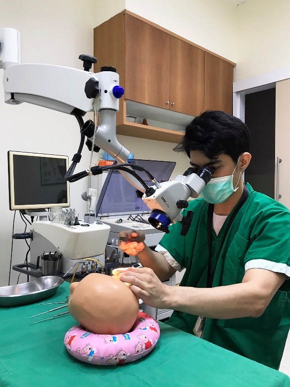
Simulated Surgical Model Design for Myringotomy and Tympanostomy Tube Insertion in Children using Medical Image Processing and 3D-Printing Technologies
DOI:
https://doi.org/10.33192/Smj.2022.79Keywords:
Myringotomy, tympanostomy tube insertion, medical image processing, 3D-print, surgical simulationAbstract
Objective: Researchers aimed to design surgical simulation models using medical image processing and 3D-printing technologies to train otolaryngologie residents with correct surgical techniques and study their skills improvement.
Materials and Methods: The models were produced for three age ranges (group A: 8-12 years old, group B: 3-7 years old, and group C: 10 months - 2 years old). Eleven residents were practiced from older to younger child models. Overall surgical time and results were evaluated to determine improvement. Both residents and specialists assessed satisfaction surveys after training.
Results: The median operational time was significantly reduced by 64.57% in model A and 50.24% in model B (p < 0.05). Operating time and surgical skills improved in order from models A, B, and C. Model C showed the most improvement with correct operational techniques in myringotomy incision (66.7%, p = 0.003) and tympanostomy tube insertion (48.5%, p = 0.011). Residents’ and specialists’ satisfaction assessments exhibited prominent satisfaction results with surgical simulation model training.
Conclusion: Surgical simulation models training enhanced residencies’ confidence and improved correct surgical techniques. Residencies can gradually practice skills from fundamental to more complicated techniques in younger child model where symptom occurs.
References
Ito M, Takahashi H, Iino Y, Kojima H, Hashimoto S, Kamide Y, et al. Clinical practice guidelines for the diagnosis and management of otitis media with effusion (OME) in children in Japan, 2015. Auris Nasus Larynx. 2017;44(5):501-8.
Wallace IF, Berkman ND, Lohr KN, Harrison MF, Kimple AJ, Steiner MJ. Surgical treatments for otitis media with effusion: a systematic review. Pediatrics. 2014;133(2):296-311.
Escamilla Y, Aguilà AF, Saiz JM, Rosell R, Vivancos J, Cardesín A. Tympanostomy tube emplacement in children with secretory otitis media: analysis of effects and complications. Acta Otorrinolaringologica (English Edition). 2009;60(2):84-9.
Rosenfeld RM, Schwartz SR, Pynnonen MA, Tunkel DE, Hussey HM, Fichera JS, et al. Clinical practice guideline: Tympanostomy tubes in children. Otolaryngol Head Neck Surg. 2013;149(1 Suppl):S1-35.
Rosenfeld RM, Shin JJ, Schwartz SR, Coggins R, Gagnon L, Hackell JM, et al. Clinical Practice Guideline: Otitis Media with Effusion (Update). Otolaryngol Head Neck Surg. 2016;154( 1 Suppl):S1-S41.
Ungkanont K. Long-term Outcome of the Management of Otitis Media with Effusion in Children with and Without Cleft Palate Using the House-brand Polyethylene Ventilation Tube Insertion. Siriraj Med J. 2021;73(4):245-51.
Suvarnsit K, Chantarawiwat T, Prakairungthong S, Limviriyakul S, Atipas S, Pitathawatchai P. Granular Myringitis Treatment at Siriraj Hospital. Siriraj Med J. 2020;72(6):502-7.
Hong P, Webb AN, Corsten G, Balderston J, Haworth R, Ritchie K, et al. An anatomically sound surgical simulation model for myringotomy and tympanostomy tube insertion. Int J Pediatr Otorhinolaryngol. 2014;78(3):522-9.
Molin N, Chiu J, Liba B, Isaacson G. Low cost, easy-toreplicate myringotomy tube insertion simulation model. Int J Pediatr Otorhinolaryngol. 2020;131(109847):1-4.
Wiet GJ, Deutsch ES, Malekzadeh S, Onwuka AJ, Callender NW, Seidman MD, et al. SimTube: A National Simulation Training and Research Project. Otolaryngol Head Neck Surg. 2020;163(3):522-30.
Barber SR, Kozin ED, Dedmon M, Lin BM, Lee K, Sinha S, et al. 3D-printed pediatric endoscopic ear surgery simulator for surgical training. Int J Pediatr Otorhinolaryngol. 2016;90:113-8.
Kuru I, Maier H, Muller M, Lenarz T, Lueth TC. A 3D-printed functioning anatomical human middle ear model. Hear Res. 2016;340:204-13.
Sparks D, Kavanagh KR, Vargas JA, Valdez TA. 3D printed myringotomy and tube simulation as an introduction to otolaryngology for medical students. Int J Pediatr Otorhinolaryngol. 2020;128(109730):1-4.
Volsky PG, Hughley BB, Peirce SM, Kesser BW. Construct validity of a simulator for myringotomy with ventilation tube insertion. Otolaryngol Head Neck Surg. 2009;141(5):603-8 e1.
Hwang JJ, Jung YH, Cho BH. The need for DICOM encapsulation of 3D scanning STL data. Imaging Sci Dent. 2018;48(4):301-2.
Kamio T, Suzuki M, Asaumi R, Kawai T. DICOM segmentation and STL creation for 3D printing: a process and software package comparison for osseous anatomy. 3D Print Med. 2020;6(1):1-12.
Edelmers E, Kazoka D, Pilmane M. Creation of Anatomically Correct and Optimized for 3D Printing Human Bones Models. Applied System Innovation. 2021;4(3):1-14.
Kumar Sharma R, Dhiman S, Negi S. Basics and applications of rapid prototyping medical models. Rapid Prototyping Journal. 2014;20(3):256-67.
Rengier F, Mehndiratta A, von Tengg-Kobligk H, Zechmann CM, Unterhinninghofen R, Kauczor HU, et al. 3D printing based on imaging data: review of medical applications. Int J Comput Assist Radiol Surg. 2010;5(4):335-41.
Ho AK, Alsaffar H, Doyle PC, Ladak HM, Agrawal SK. Virtual reality myringotomy simulation with real-time deformation: development and validity testing. Laryngoscope. 2012;122(8):1844-51.
Huang C, Cheng H, Bureau Y, Agrawal SK, Ladak HM. Face and content validity of a virtual-reality simulator for myringotomy with tube placement. J Otolaryngol Head Neck Surg. 2015;44(40):1-8.
Huang C, Cheng H, Bureau Y, Ladak HM, Agrawal SK. Automated Metrics in a Virtual-Reality Myringotomy Simulator: Development and Construct Validity. Otol Neurotol. 2018;39(7):e601-e8.
Vaitaitis VJ, Dunham ME, Kwon YC, Mayer WC, Evans AK, Baker AJ, et al. A Surgical Simulator for Tympanostomy Tube Insertion Incorporating Capacitive Sensing Technology to Track Instrument Placement. Otolaryngol Head Neck Surg. 2020;162(3):343-5.

Published
How to Cite
Issue
Section
License

This work is licensed under a Creative Commons Attribution-NonCommercial-NoDerivatives 4.0 International License.
Authors who publish with this journal agree to the following conditions:
Copyright Transfer
In submitting a manuscript, the authors acknowledge that the work will become the copyrighted property of Siriraj Medical Journal upon publication.
License
Articles are licensed under a Creative Commons Attribution-NonCommercial-NoDerivatives 4.0 International License (CC BY-NC-ND 4.0). This license allows for the sharing of the work for non-commercial purposes with proper attribution to the authors and the journal. However, it does not permit modifications or the creation of derivative works.
Sharing and Access
Authors are encouraged to share their article on their personal or institutional websites and through other non-commercial platforms. Doing so can increase readership and citations.


