Three-dimensional Kinematic Analysis and Muscle Activation of the Upper Extremity in Ruesi Dutton Exercises
DOI:
https://doi.org/10.33192/Smj.2022.85Keywords:
Ruesi-Dutton, Hermit Doing Body Contortion, biomechanics, upper extremityAbstract
Objective: To investigate 3-D upper extremity joint angles and muscle activities in selected Ruesi-Dutton exercises.
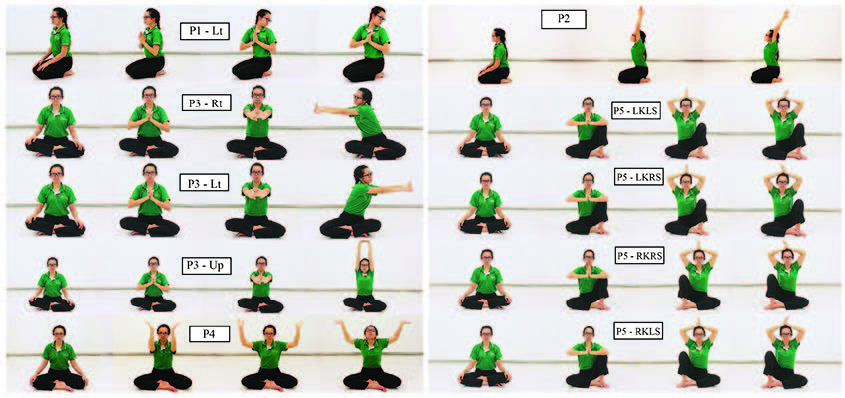
Material and Methods: Twenty-six healthy participants (mean age of 25.65, mean height of 165.08 cm, and mean weight of 56.69 Kg) volunteered to take part in this study. 3-D motion analysis consisted of eight cameras synchronized with a wireless electromyography (EMG) system to collect kinematic data and muscle activity. Participants performed five postures, including the Kae Lom Kho Mue posture, Kae Puat Thong Kae Kho Thao posture, Kae Kiat posture, Kae Puat Thong Sabak Chom posture, and Kae Lom Puat Sisa. The upper extremity joint angles and range of motion (ROM) and EMG were analyzed.
Results: Most postures were in the normal range of motion. The percentage of MVIC was more than 1% and the Trapezius muscle is the most active in all postures.
Conclusion: The data in this research is useful to help select the correct posture and exercise for a specific condition.
References
The Fine Arts Department. Samut Phap Khlong Ruesi Datton. Bangkok: Amarin Printing and Publishing, 2551.
Somdet Phraborom Wong Thoe Kromphraya Damrong Ra Chanu Phap. “Ruesi Datton”. Nithan Borankhadi, 10th ed. Bangkok: Khasem Ban Na Kit, 2503.
The Office of Royal Society. Photchananukrom Chabap Ratchabandittayasathan B.E. 2554 Chaloemphrakiat PhraBatSomdetPhrachaoyuhua Nueang Nai Okat Phraratchaphithi Maha Mongkhon Chaloemphrachonphansa anniversary 7th. Bangkok: Sirivatana Interprint, 2556.
Ayurved Thamrong school, Center of Applied Thai Traditional Medicine, Faculty of Siriraj Medicine Hospital, Mahidol university. Kaiborihan Baep Ruesi Datton. Vol 1. Bangkok: Suppha Wanit Publishing, 2554.
Butdapan P, Narajeenarone K, Apichartvorakit A, Kade S, Jansomsarit S, Jamjuntra P, et al. The Effect of the Posture of the “Hermit Doing Body Contortion” on Relief of Shoulder and Scapular Pain Caused by Chronic Myofascial Pain Syndrome: A Randomized, Parallel Group, Controlled Trial. Siriraj Med J. 2016;68:350-7.
Tanasugarn L, Natearpha P, Kongsakon R, Chaosaowapa M, Choatwongwachira W, Seanglaw D, et al. Physical effects and cognitive function after exercising “Rue-si-dad-ton” (Exercise using the posture of the hermit doing body contortion): A randomized controlled pilot trial. J Med Assoc Thai. 2015;98(3):306-13.
Ngowsiri K, Tanmahasamut P, Sukonthasab S. Rusie Dutton traditional Thai exercise promotes health related physical fitness and quality of life in menopausal women. Complement Ther Clin Pract. 2014;20(3):164-71.
Inokuchi H, Tojima M, Mano H, Ishikawa Y, Ogata N, Haga N. Neck range of motion measurements using a new threedimensional motion analysis system: validity and repeatability. Eur Spine J. 2015: 2807-15.
SENIAM Project [Internet]. Netherland: Roessingh Research and Development; 2006 [cited 2019 Mar 9]. Available from: http://seniam.org/
Konrad P. The ABC of EMG-A practical introduction to Kinesiological Electromyography. U.S.A. Noraxon Inc., 2006.
Vicon Motion Systems Limited. Plug-in Gait Reference Guide [Internet]. 2017 [cited 2021 Dec 29]. Available from: https://docs.vicon.com/
Boone DC, Azen SP. Normal range of motion of joints in male subjects. JBJS. 1979;61(5):756-9.
Youdas JW, Garrett TR, Suman VJ, Bogard CL, Hallman HO, Carey JR. Normal range of motion of the cervical spine: an initial goniometric study. Phys Ther. 1992;72(11):770-80.
Barnes CJ, Van Steyn SJ, Fischer RA. The effects of age, sex, and shoulder dominance on range of motion of the shoulder. J Shoulder Elbow Surg. 2001;10(3):242-6.
Rundquist PJ, Obrecht C, Woodruff L. Three-dimensional shoulder kinematics to complete activities of daily living. Am J Phys Med Rehabil. 2009;88(8):623-9.
Namdari S, Yagnik G, Ebaugh DD, Nagda S, Ramsey ML, Williams Jr GR, et al. Defining functional shoulder range of motion for activities of daily living. J Shoulder Elbow Surg. 2012;21(9):1177-83.
Ryu J, Cooney III WP, Askew LJ, An KN, Chao EY. Functional ranges of motion of the wrist joint. J Hand Surg. 1991;16(3):409-19.
Akalin E, El Ö, Peker Ö, Senocak Ö, Tamci S, Gülbahar S, et al. Treatment of carpal tunnel syndrome with nerve and tendon gliding exercises. Am J Phys Med Rehabil. 2002;81(2):108-13.
Castro WH, Sautmann A, Schilgen M, Sautmann M. Noninvasive three-dimensional analysis of cervical spine motion in normal subjects in relation to age and sex: an experimental examination. Spine. 2000;25(4):443-9.
Rundquist PJ, Anderson DD, Guanche CA, Ludewig PM. Shoulder kinematics in subjects with frozen shoulder. Arch Phys Med Rehabil. 2003;84(10):1473-9.
Headaches disorder [Internet]. Switzerland: World Health Organization; 2016 [cited 2022 Jan 14]. Available from: https://www.who.int/news-room/fact-sheets/detail/headache-disorders.
Prateepavanich P. Myofascial pain syndrome ca. In: Prateepavanich P, Chaudakshetrin P, editors. Myofascial pain syndrome: a common problem in clinical practice. 1st ed. Bangkok: Amarin Printing and Publishing; 2542.p.273-320.
Prateepavanich P. Subscapularis Muscle. In: Prateepavanich P, Chaudakshetrin P, editors. Myofascial pain syndrome: a common problem in clinical practice. 1st ed. Bangkok: Amarin Printing and Publishing; 2542.p.388-91.
Chopp-Hurley JN, Prophet C, Thistle B, Pollice J, Maly MR. Scapular muscle activity during static yoga postures. J Orthop Sports Phys Ther. 2018;48(6):504-9.
Govindaraj R, Karmani S, Varambally S, Gangadhar BN. Yoga and physical exercise–a review and comparison. Int Rev Psychiatry 2016;28(3):242-53.
Lauver JD, Cayot TE, Scheuermann BW. Influence of bench angle on upper extremity muscular activation during bench press exercise. Eur J Sport Sci. 2016;16(3):309-16.
Scott R, Yang HS, James CR, Sawyer SF, Sizer Jr PS. Volitional preemptive abdominal contraction and upper extremity muscle latencies during D1 flexion and scaption shoulder exercises. J Athl Train. 2018;53(12):1181-9.

Published
How to Cite
Issue
Section
License

This work is licensed under a Creative Commons Attribution-NonCommercial-NoDerivatives 4.0 International License.
Authors who publish with this journal agree to the following conditions:
Copyright Transfer
In submitting a manuscript, the authors acknowledge that the work will become the copyrighted property of Siriraj Medical Journal upon publication.
License
Articles are licensed under a Creative Commons Attribution-NonCommercial-NoDerivatives 4.0 International License (CC BY-NC-ND 4.0). This license allows for the sharing of the work for non-commercial purposes with proper attribution to the authors and the journal. However, it does not permit modifications or the creation of derivative works.
Sharing and Access
Authors are encouraged to share their article on their personal or institutional websites and through other non-commercial platforms. Doing so can increase readership and citations.


