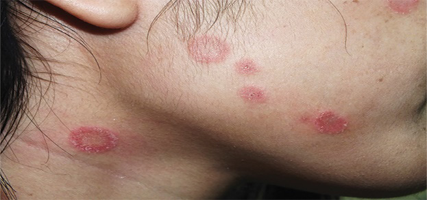
Human-pet Relationship, Pet Abandonment, and Clinical Correlation for Patients Infected with Dermatophytosis of the Glabrous Skin
DOI:
https://doi.org/10.33192/smj.v75i2.260745Keywords:
Dermatophyte, animals, Trichophyton, Microsporum, petAbstract
Objective: The study on human-pet relationship and pet abandonment among dermatophytosis patients is limited. This study aims to review these correlations.
Materials & Methods: A two-year retrospective cross-sectional study was conducted. Case record forms were reviewed for clinical manifestations, fungal identification, human-pet relationships, and changes in the relationships after dermatophytosis diagnosis.
Results: A total of 230 dermatophytosis patients from the Dermatology outpatient clinic, Siriraj Hospital, were included. The mean age was 41.9 ± 19.1 years and 51.3% were female. Among 170 cases with positive fungal culture, zoophilic dermatophytosis from M. canis infection was identified in 15.9% which was predominately found in females and manifested as shorter duration of onset, and higher involvement on exposed areas when compared to anthropophilic dermatophytosis. Most (71%%) of patients with M. canis infection classified themselves as pet-lovers. The relationship with pets had changed after the dermatophytosis diagnosis in 41% of them which was statistically different from 8.8% in non-pet lovers (P = 0.001). The overall pet abandonment rate was 26.6%. The abandonment rate was 40.9% among non-pet lovers, while 30.6% was reported among pet lovers.
Conclusion: Zoophilic M. canis infection was associated with rapid onset and on predominant-exposed areas. Some pets could be asymptomatic, so identification of the reservoirs of dermatophytosis is important in the treatment process and helps prevent future recurrence. Paying attention to human-pet relationships and pet abandonment is critical. Knowledge about dermatophytosis transmission, and appropriate pet management should be advised to decrease abandonment.
References
Pires CA, Cruz NF, Lobato AM, Sousa PO, Carneiro FR, Mendes AM. Clinical, epidemiological, and therapeutic profile of dermatophytosis. An Bras Dermatol 2014;89(2):259-64.
Craddock LN, SM S. Superficial Fungal Infection. In: Kang S, Amagai M, Bruckner AL, Enk AH, Margolis DJ, McMichael AJ, et al., eds. Fitzpatrick's Dermatology, 9th ed: McGraw-Hill, 2019. p. 2927.
Weitzman I, Summerbell RC. The dermatophytes. Clin Microbiol Rev 1995;8(2):240-59.
Bunyaratavej S, Kiratiwongwan R, Limphoka P, Lertrujiwanit K, Leeyaphan C. Pattern Recognition using Morphologies of Anthropophilic and Zoophilic Dermatophytosis Lesions: Comparison between Final-Year Medical Students and Dermatology Residents. Siriraj Med J 2020;72(6):488-91.
Aghamirian MR, Ghiasian SA. Dermatophytoses in outpatients attending the Dermatology Center of Avicenna Hospital in Qazvin, Iran. Mycoses 2008;51(2):155-60.
Seebacher C, Bouchara JP, Mignon B. Updates on the epidemiology of dermatophyte infections. Mycopathologia 2008;166(5-6):335-52.
Cai W, Lu C, Li X, Zhang J, Zhan P, Xi L, et al. Epidemiology of Superficial Fungal Infections in Guangdong, Southern China: A Retrospective Study from 2004 to 2014. Mycopathologia 2016;181(5-6):387-95.
Rashidian S, Falahati M, Kordbacheh P, Mahmoudi M, Safara M, Sadeghi Tafti H, et al. A study on etiologic agents and clinical manifestations of dermatophytosis in Yazd, Iran. Curr Med Mycol 2015;1(4):20-5.
Chermette R, Ferreiro L, Guillot J. Dermatophytoses in animals. Mycopathologia 2008;166(5-6):385-405.
Havlickova B, Czaika VA, Friedrich M. Epidemiological trends in skin mycoses worldwide. Mycoses 2008;51 Suppl 4:2-15.
Bunyaratavej S, Limphoka P, Kiratiwongwan R, Leeyaphan C. Survey of skin and nail fungal infections by subject age among thai adults and the etiological organisms. Southeast Asian J Trop Med Public Health SE 2019;50(6):1118-31.
Nenoff P, Handrick W, Krüger C, Vissiennon T, Wichmann K, Gräser Y, et al. Dermatomykosen durch Haus- und Nutztiere. Der Hautarzt 2012;63(11):848-58.
Cafarchia C, Romito D, Sasanelli M, Lia R, Capelli G, Otranto D. The epidemiology of canine and feline dermatophytoses in southern Italy. Mycoses 2004;47(11-12):508-13.
Halsby KD, Walsh AL, Campbell C, Hewitt K, Morgan D. Healthy animals, healthy people: zoonosis risk from animal contact in pet shops, a systematic review of the literature. PLoS One 2014;9(2):e89309.
Katoh T, Maruyama R, Nishioka K, Sano T. Tinea corporis due to Microsporum canis from an asymptomatic dog. J Dermatol 1991;18(6):356-9.
Mancianti F, Nardoni S, Corazza M, D'Achille P, Ponticelli C. Environmental detection of Microsporum canis arthrospores in the households of infected cats and dogs. J Feline Med Surg 2003;5(6):323-8.
Shiraki Y, Hiruma M, Matsuba Y, Kano R, Makimura K, Ikeda S, et al. A case of tinea corporis caused by Arthroderma benhamiae (teleomorph of Tinea mentagrophytes) in a pet shop employee. J Am Acad Dermatol 2006;55(1):153-4.
Stull JW, Peregrine AS, Sargeant JM, Weese JS. Household knowledge, attitudes and practices related to pet contact and associated zoonoses in Ontario, Canada. BMC Public Health 2012;12(1):553.
Kobwanthanakun W, Bunyaratavej S, Leeyaphan C. Tinea corporis from Microsporum canis: A case report in 2 patients from 1 asymptomatic feline. Thai J Dermatol 2018;34(4):299-304.
Iorio R, Cafarchia C, Capelli G, Fasciocco D, Otranto D, Giangaspero A. Dermatophytoses in cats and humans in central Italy: epidemiological aspects. Mycoses 2007;50(6):491-5.
Bunyaratavej S LP, Kiratiwongwan R, Leeyaphan C. Comparison of patient and clinical differences between superficial skin infections due to Microsporum canis and Trichophyton rubrum. Southeast Asian J Trop Med Public Health SE 2020;51(1).
Limphoka P, Bunyaratavej S, Leeyaphan C. Fingernail onychomycosis caused by Microsporum canis in a teenager. Pediatr Dermatol 2021;38(2):524-5.
Cafarchia C, Romito D, Capelli G, Guillot J, Otranto D. Isolation of Microsporum canis from the hair coat of pet dogs and cats belonging to owners diagnosed with M. canis tinea corporis. Vet Dermatol 2006;17(5):327-31.
Moriello KA, Coyner K, Paterson S, Mignon B. Diagnosis and treatment of dermatophytosis in dogs and cats.: Clinical Consensus Guidelines of the World Association for Veterinary Dermatology. Vet Dermatol 2017;28(3):266-e68.
Ali-Shtayeh MS, Yaish S, Jamous RM, Arda H, Husein EI. Updating the epidemiology of dermatophyte infections in Palestine with special reference to concomitant dermatophytosis. J Mycol Med 2015;25(2):116-22.
Bunyaratavej S, Kiratiwongwan R, Suphatsathienkul P, Munprom K, Matthapan L, Supcharoenkul S, et al. Effect of Different Shampoos and Contact Time on Microsporum canis Infected Hair: In vitro Model Study. Thai J Dermatol 2020;36(4):150-6.

Published
How to Cite
Issue
Section
Categories
License

This work is licensed under a Creative Commons Attribution-NonCommercial-NoDerivatives 4.0 International License.
Authors who publish with this journal agree to the following conditions:
Copyright Transfer
In submitting a manuscript, the authors acknowledge that the work will become the copyrighted property of Siriraj Medical Journal upon publication.
License
Articles are licensed under a Creative Commons Attribution-NonCommercial-NoDerivatives 4.0 International License (CC BY-NC-ND 4.0). This license allows for the sharing of the work for non-commercial purposes with proper attribution to the authors and the journal. However, it does not permit modifications or the creation of derivative works.
Sharing and Access
Authors are encouraged to share their article on their personal or institutional websites and through other non-commercial platforms. Doing so can increase readership and citations.


