Prevalence and Trend of Photodermatoses in Thailand: A 16-year Retrospective Study at Siriraj Hospital
DOI:
https://doi.org/10.33192/smj.v75i2.260748Keywords:
Photodermatoses, drug-induced photosensitivity, immunologically-mediated photodermatoses, photo-aggravated dermatoses, genophotodermatoses, polymorphous light eruptionAbstract
Objective: Photodermatoses are a group of cutaneous disorders with abnormal reactions triggered by exposure to sunlight. Previous studies reported varying photodermatoses prevalence in Caucasians and African-Americans; however, it was seldom reported in the Asian population. The aim of our study was to determine the prevalence, clinical characteristics and trend of photodermatoses in Thailand.
Materials & Methods: A retrospective chart review was performed at the Faculty of Medicine Siriraj Hospital, Mahidol University using diagnoses from the International Classification of Disease (ICD), Tenth Revisions codes, between January 2005 and September 2021.
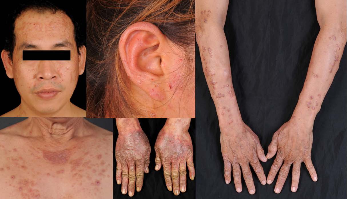
Results: A total of 561 patients with definite diagnosis of photodermatoses were identified. The prevalence of photodermatoses in the outpatient dermatology clinic was 3 cases per 1,000. The most common photodermatoses was chemical and drug-induced photosensitivity (39.4%), followed by immunologically-mediated photodermatoses (30.1%), photo-aggravated dermatoses (29.4%) and genophotodermatoses (1.1%). Overall phototesting was performed in 276 cases (49.2%). In our study, some photodermatoses had unique clinical characteristics including pinpoint popular variant of polymorphous like eruption and adult-onset actinic prurigo. Over 16 years, the trend of patients being diagnosed with photodermatoses has continued to rise gradually with an increment of 1.67 times.
Conclusion: Photodermatoses are uncommon in Thailand. Some photodermatoses have distinctive clinical features in Asian populations. The trend of photodermatoses in Thailand is continually rising, reflecting an increase in physicians’ awareness and knowledge of these cutaneous conditions.
References
Gutierrez D, Gaulding JV, Motta Beltran AF, Lim HW, Pritchett EN. Photodermatoses in skin of colour. J Eur Acad Dermatol Venereol. 2018;32(11):1879-86.
Bylaite M, Grigaitiene J, Lapinskaite GS. Photodermatoses: classification, evaluation and management. Br J Dermatol. 2009;161 Suppl 3:61-8.
Choi D, Kannan S, Lim HW. Evaluation of patients with photodermatoses. Dermatol Clin. 2014;32(3):267-75.
Hönigsmann H, Hojyo-Tomoka MT. Polymorphous light eruption, hydroa vacciniforme, and actinic prurigo. Photodermatol: CRC Press; 2007.p.149-68.
Deng D, Hang Y, Chen H, Li H. Prevalence of photodermatosis in four regions at different altitudes in Yunnan province, China. J Dermatol. 2006;33(8):537-40.
Wong SN, Khoo LS. Analysis of photodermatoses seen in a predominantly Asian population at a photodermatology clinic in Singapore. Photodermatol Photoimmunol Photomed. 2005;21(1):40-4.
Nakamura M, Henderson M, Jacobsen G, Lim HW. Comparison of photodermatoses in African-Americans and Caucasians: a follow-up study. Photodermatol Photoimmunol Photomed. 2014;30(5):231-6.
Kerr HA, Lim HW. Photodermatoses in African Americans: a retrospective analysis of 135 patients over a 7-year period. J Am Acad Dermatol. 2007;57(4):638-43.
Hamel R, Mohammad TF, Chahine A, Joselow A, Vick G, Radosta S, et al. Comparison of racial distribution of photodermatoses in USA academic dermatology clinics: A multicenter retrospective analysis of 1,080 patients over a 10-year period. Photodermatol Photoimmunol Photomed. 2020;36(3):233-40.
Ibbotson S, Dawe R. Cutaneous Photosensitivity Diseases. In: Griffiths CEM, Barker J, Bleiker T, Chalmers R, Creamer D, editors. In Rook’s Textbook of Dermatology. 9th ed. John Wiley & Sons; 2016.p.127.1-127.35.
Dawe RS. Prevalences of chronic photodermatoses in Scotland. Photodermatol Photoimmunol Photomed. 2009;25(1):59-60.
Khoo SW, Tay YK, Tham SN. Photodermatoses in a Singapore skin referral centre. Clin Exp Dermatol. 1996;21(4):263-8.
Blakely KM, Drucker AM, Rosen CF. Drug-Induced Photosensitivity-An Update: Culprit Drugs, Prevention and Management. Drug Saf. 2019;42(7):827-47.
Fotiades J, Soter NA, Lim HW. Results of evaluation of 203 patients for photosensitivity in a 7.3-year period. J Am Acad Dermatol. 1995;33(4):597-602.
Akaraphanth R, Sindhavananda J, Gritiyarangsan P. Adult-onset actinic prurigo in Thailand. Photodermatol Photoimmunol Photomed. 2007;23(6):234-7.
Lestarini D, Khoo LS, Goh CL. The clinical features and management of actinic prurigo: A retrospective study. Photodermatol Photoimmunol Photomed 1999;15:183-7.
Gruber-Wackernagel A, Wolf P. Polymorphic Light Eruption. In: Kang S, Amagai M, Bruckner AL, et al., eds. Fitzpatrick's Dermatology, 9th ed. New York: McGraw-Hill Education; 2019.
Ellenbogen E, Wesselmann U, Hofmann SC, Lehmann P. Photosensitive atopic dermatitis-a neglected subset: Clinical, laboratory, histological and photobiological workup. J Eur Acad Dermatol Venereol. 2016;30(2):270-5.
Lehmann AR, McGibbon D, Stefanini M. Xeroderma pigmentosum. Orphanet J Rare Dis. 2011;6:70.
Nassan H, Dawe RS, Moseley H, Ibbotson SH. A review of photodiagnostic investigations over 26 years: experience of the National Scottish Photobiology Service (1989-2015). J R Coll Physicians Edinb. 2017;47(4):345-50.

Published
How to Cite
Issue
Section
Categories
License

This work is licensed under a Creative Commons Attribution-NonCommercial-NoDerivatives 4.0 International License.
Authors who publish with this journal agree to the following conditions:
Copyright Transfer
In submitting a manuscript, the authors acknowledge that the work will become the copyrighted property of Siriraj Medical Journal upon publication.
License
Articles are licensed under a Creative Commons Attribution-NonCommercial-NoDerivatives 4.0 International License (CC BY-NC-ND 4.0). This license allows for the sharing of the work for non-commercial purposes with proper attribution to the authors and the journal. However, it does not permit modifications or the creation of derivative works.
Sharing and Access
Authors are encouraged to share their article on their personal or institutional websites and through other non-commercial platforms. Doing so can increase readership and citations.


