Evaluation of Hair Follicle Counts of Occipital Scalp Biopsies from Male Hair Transplant Patients in Thailand
DOI:
https://doi.org/10.33192/smj.v75i2.260752Keywords:
Hair counts, hair density, occipital area, androgenetic alopeciaAbstract
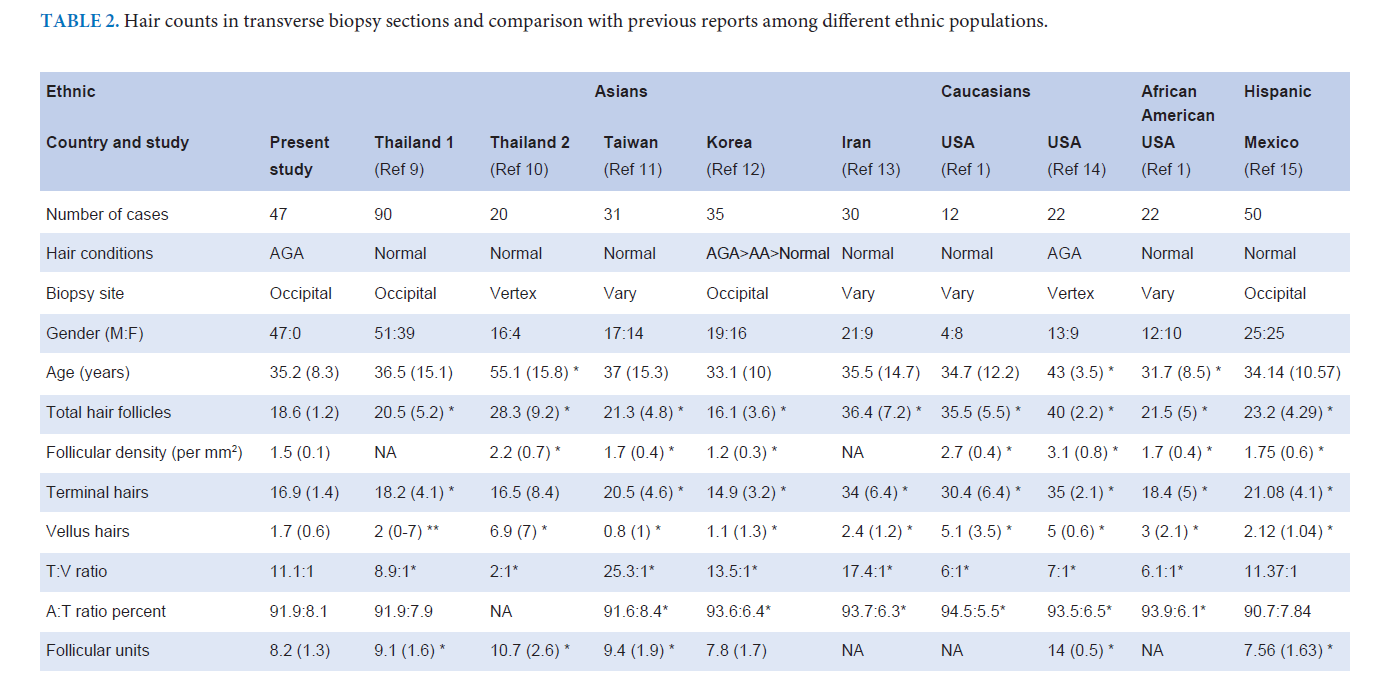
Objective: To evaluate the average hair follicle count from the occipital scalp of Thai males with AGA who were candidates for hair transplantation.
Materials & Methods: A cross-sectional study of 47 male with AGA undergoing hair transplantation surgery was conducted. The 4-mm punch biopsies from the occipital scalp were evaluated for hair count parameters. The results were compared to prior studies.
Results: The average counts of total hair follicles and the density of hair follicle per square millimeter were 18.6±1.2, and 1.5±0.1, respectively. The terminal-to-vellus ratio was 11.1, and the percent ratio of anagen-to-telogen ratio was 91.9:8.1. The hair count number is significantly lower than other ethnicities including Thais in general population (P < 0.001), but greater than the Thai males with AGA in the previous study. (P < 0.001).
Conclusion: Our study showed a lower average hair density as compared to the other normal Asian population. The total hair count in the occipital area from this study is less when compared to the previous studies conducted in Thai normal controls but higher than those with more advanced AGA. This result supported the evidence of hormonal effect involving the occipital scalp of male AGA.
References
Sperling LC. Hair density in African Americans. Arch Dermatol. 1999;135(6):656-8.
Sawaya ME, Price VH. Different levels of 5alpha-reductase type I and II, aromatase, and androgen receptor in hair follicles of women and men with androgenetic alopecia. J Invest Dermatol. 1997; 109:296-300.
Ekmekci TR, Sakiz D, Koslu A. Occipital involvement in female pattern hair loss: histopathological evidences. J Eur Acad Dermatol Venereol. 2010;24:299-301.
Khunkhet S, Chanprapaph K, Rutnin S, Suchonwanit P. Histopathological Evidence of Occipital Involvement in Male Androgenetic Alopecia. Front Med (Lausanne). 2021;22:790597.
Watanabe-Okada E, Amagai M, Ohyama M. Histopathological investigation of clinically non-affected perilesional scalp in alopecias detected unexpected spread of disease activities. J Dermatol. 2014;41:802-7.
Park JH, Park JM, Kim NR, Manonukul K. Hair diameter evaluation in different regions of the safe donor area in Asian populations. Int J Dermatol. 2017;56:784-7.
Visessiri Y, Pakornphadungsit K, Leerunyakul K, Rutnin S, Srisont S, Suchonwanit P. The study of hair follicle counts from scalp histopathology in the Thai population. Int J Dermatol. 2020;59:978-81.
Yaprohm P, Manonukul J, Sontichai V, Pooliam J, Srettabunjong S. Hair follicle counts in Thai population: a study on the vertex scalp area. J Med Assoc Thai. 2013;96:1578-82.
Ko JH, Huang YH, Kuo TT. Hair counts from normal scalp biopsy in Taiwan. Dermatol Surg. 2012;38:1516-20.
Lee HJ, Ha SJ, Lee JH, Kim JW, Kim HO, Whiting DA. Hair counts from scalp biopsy specimens in Asians. J Am Acad Dermatol. 2002;46:218-21.
Aslani FS, Dastgheib L, Banihashemi BM. Hair counts in scalp biopsy of males and females with androgenetic alopecia compared with normal subjects. J Cutan Pathol. 2009; 36:734-9.
Whiting DA. Diagnostic and predictive value of horizontal sections of scalp biopsy specimens in male pattern androgenetic alopecia. J Am Acad Dermatol. 1993;28:755-63.
Martínez-Luna E, Rodríguez-Lobato E, Vázquez-Velo JA, Cuevas-González JC, Martínez Velasco MA, Toussaint Caire S. Quantification of hair follicles in the scalp in Mexican Mestizo population. Skin Appendage Disord. 2018;5:27-31.
Palo S, Biligi DS. Utility of horizontal and vertical sections of scalp biopsies in various forms of primary alopecias. J Lab Physicians. 2018;10:95-100.
Solomon AR. The transversely sectioned scalp biopsy specimen: the technique and an algorithm for its use in the diagnosis of alopecia. Adv Dermatol. 1994;9:127-57.

Published
How to Cite
Issue
Section
Categories
License

This work is licensed under a Creative Commons Attribution-NonCommercial-NoDerivatives 4.0 International License.
Authors who publish with this journal agree to the following conditions:
Copyright Transfer
In submitting a manuscript, the authors acknowledge that the work will become the copyrighted property of Siriraj Medical Journal upon publication.
License
Articles are licensed under a Creative Commons Attribution-NonCommercial-NoDerivatives 4.0 International License (CC BY-NC-ND 4.0). This license allows for the sharing of the work for non-commercial purposes with proper attribution to the authors and the journal. However, it does not permit modifications or the creation of derivative works.
Sharing and Access
Authors are encouraged to share their article on their personal or institutional websites and through other non-commercial platforms. Doing so can increase readership and citations.


