
Predicting Progression to Hypervascular HCC in Hypovascular Hypointense Nodules in Gadoxetic Acid-enhanced MR Images in Patients with Chronic Liver Disease
DOI:
https://doi.org/10.33192/smj.v75i9.262021Keywords:
Gadoxetic acid-enhanced MRI, hypovascular hypointense nodule in hepatobiliary phase, HCC imagingAbstract
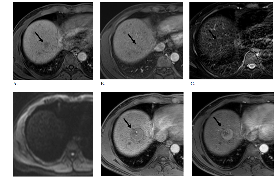
Objective: To identify patient characteristics and MR imaging features of hypovascular hypointense nodules in the hepatobiliary phase (HBP) of gadoxetic acid-enhanced MR imaging in patients with chronic liver disease associated with progression to hypervascular hepatocellular carcinoma (HCC).
Materials and Methods: The institutional review board approved this retrospective review of 40 patients with 60 hypovascular hypointense nodules in the HBP of gadoxetic acid-enhanced MR imaging. Univariate and multivariate Cox regression analyses for hypervascular HCC development were used to define variables, including initial nodule size, cause of cirrhosis, history of locoregional therapy of HCC, fat-containing, signal intensity on T1W, T2W, portal and equilibrium phases of dynamic phase, and DW images. The cumulative percentage incidence of hypervascularity and growth rate were calculated using the receiver operating characteristic (ROC) curve.
Results: The prevalence of progression to hypervascular HCC was 45% (27 out of 60). The Multivariable Cox analysis of developing hypervascularization was an initial nodule diameter more than 1 cm. (P=0.027; HR 2.52; 95% CI: 1.11,5.74) The mean growth rate was significantly higher in subsequent hypervascular nodules than in those without hypervascularization (P < 0.001). The cumulative risk incidence of hypervascularization at 3, 6, 12, 24 months was 5%, 20%, 35%, 44 %, respectively.
Conclusion: An initial nodule diameter of more than 1 cm and nodules with higher growth rates are significant predictive factors for hypervascular transformation of hypovascular hypointense nodules in the HBP of gadoxetic acid-enhanced MR imaging.
References
Sakamoto M, Hirohashi S, Shimosato Y. Early stages of multistep hepatocarcinogenesis: adenomatous hyperplasia and early hepatocellular carcinoma. Hum Pathol. 1991;22(2):172–8.
Takayama T, Makuuchi M, Hirohashi S, Sakamoto M, Okazaki N, Takayasu K, et al. Malignant transformation of adenomatous hyperplasia to hepatocellular carcinoma. Lancet. 1990;336(8724):1150–3.
Kudo M. Multistep human hepatocarcinogenesis: correlation of imaging with pathology. J Gastroenterol. 2009;44(Suppl 19):112–8.
Marrero JA, Kulik LM, Sirlin CB, Zhu AX, Finn RS, Abecassis MM, et al. Diagnosis, staging, and management of hepatocellular carcinoma: 2018 practice guidance by the american association for the study of liver diseases. Hepatology. 2018;68(2):723-50.
EASL Clinical Practice Guidelines: Management of hepatocellular carcinoma. J Hepatol. 2018;69:182-236.
2018 Korean Liver Cancer Association-National Cancer Center Korea Practice Guidelines for the management of hepatocellular carcinoma. Korean J Radiol. 2019;20(7):1042-113.
Omata M, Cheng AL, Kokudo N, Kudo M, Lee JM, Jia J, et al. Asia-Pacific clinical practice guidelines on the management of hepatocellular carcinoma: a 2017 update. Hepatol Int. 2017;11:317-70.
Yoon SH, Lee JM, So YH, Hong SH, Kim SJ, Han JK, et al. Multiphasic MDCT enhancement pattern of hepatocellular carcinoma smaller than 3 cm in diameter: tumor size and cellular differentiation. AJR. 2009;193:482-9.
Teerasamit W, Tongdee R, Yodying J. Diagnostic Performance of Gadoxetic Acid-Enhanced MR Imaging in the Diagnosis of Hepatocellular Carcinoma in Cirrhotic Liver. J Med Assoc Thai. 2017;100(8):918-26.
Cruite I, Schroeder M, Merkle EM, Sirlin CB. Gadoxetate disodium-enhanced MRI of the liver: part 2, protocol optimization and lesion appearance in the cirrhotic liver. AJR Am J Roentgenol. 2010; 195:29-41.
Kim MJ. Current limitations and potential breakthroughs for the early diagnosis of hepatocellular carcinoma. Gut Liver. 2011;5(1):15–21.
Park HJ, Choi BI, Lee ES, Park SB, Lee JB. How to differentiate borderline hepatic nodules in hepatocarcinogenesis: Emphasis on imaging diagnosis. Liver Cancer. 2017;6:189-203.
Hayashi M, Matsui O, Ueda K, Kawamori Y, Gabata T, Kadoya M. Progression to hypervascular hepatocellular carcinoma: correlation with intranodular blood supply evaluated with CT during intraarterial injection of contrast material. Radiology. 2002;225(1):143–9.
Kumada T, Toyoda H, Tada T, Sone Y, Fujimori M, Ogawa S, et al. Evolution of hypointense hepatocellular nodules observed only in the hepatobiliary phase of gadoxetate disodium–enhanced MRI. Am J Roentgenol. 2011;197:58–63.
Motosugi U, Ichikawa T, Sano K, Sou H, Onohara K, Muhi A, et al. Outcome of hypovascular hepatic nodules revealing no gadoxetic acid uptake in patients with chronic liver disease. J Magn Reson Imaging. 2011;34:88–94.
Suh CH, Kim KW, Pyo J, Lee J, Kim SY, Park SH. Hypervascular transformation of hypovascular hypointense nodules in the hepatobiliary phase of gadoxetic acid-enhanced MRI: a systematic review and meta-analysis. AJR Am J Roentgenol. 2017; 209(4):781-9.
Kim YS, Song JS, Lee HK, Han YM. Hypovascular hypointense nodules on hepatobiliary phase without T2 hyperintensity on gadoxetic acid-enhanced MR images in patients with chronic liver disease: long-term outcomes and risk factors for hypervascular transformation. Eur Radiol. 2016; 26(10):3728-36.
Hyodo T, Murakami T, Imai Y, Okada M, Hori M, Kagawa Y, et al. Hypovascular nodules in patients with chronic liver disease: risk factors for development of hypervascular hepatocellular carcinoma. Radiology. 2013;266(2):480-90.
Lee MH, Kim SH, Park MJ, Park CK, Rhim H. Gadoxetic acid-enhanced hepatobiliary phase MRI and high b-value diffusion weighted imaging to distinguish well-differentiated hepatocellular carcinomas from benign nodules in patients with chronic liver disease. AJR Am J Roentgenol. 2011;197:W868-W75.
Kim YK, Lee WJ, Park MJ, Kim SH, Rhim H, Choi D. Hypovascular hypointense nodules on hepatobiliary phase gadoxetic acid – enhanced MR images in patients with cirrhosis: Potential of DW imaging in predicting progression to hypervascular HCC. Radiology. 2012;266(2):104-12.
Briani C, Pietropaolo MD, Marignani M, Carbonetti F, Begini P, David V, et al. Non-Hypervascular Hypointense Nodules at Gadoxetic Acid MRI: Hepatocellular Carcinoma Risk Assessment with Emphasis on the Role of Diffusion-Weighted Imaging. J Gastrointest Cancer. 2018; 49(3):302-10.
Published
How to Cite
License
Copyright (c) 2023 Siriraj Medical Journal

This work is licensed under a Creative Commons Attribution-NonCommercial-NoDerivatives 4.0 International License.
Authors who publish with this journal agree to the following conditions:
Copyright Transfer
In submitting a manuscript, the authors acknowledge that the work will become the copyrighted property of Siriraj Medical Journal upon publication.
License
Articles are licensed under a Creative Commons Attribution-NonCommercial-NoDerivatives 4.0 International License (CC BY-NC-ND 4.0). This license allows for the sharing of the work for non-commercial purposes with proper attribution to the authors and the journal. However, it does not permit modifications or the creation of derivative works.
Sharing and Access
Authors are encouraged to share their article on their personal or institutional websites and through other non-commercial platforms. Doing so can increase readership and citations.


