
A Case Series and Systematic Review: Results of Surgical Management of Glaucoma Drainage Device Tube Exposure
DOI:
https://doi.org/10.33192/smj.v76i12.270377Keywords:
Glaucoma drainage device, Glaucoma tube, Glaucoma shunt, Expose, TreatmentAbstract
Objective: To present a case series of patients who underwent surgical repair for glaucoma drainage device (GDD) tube exposure and conduct a systematic review to analyze results of various surgical techniques.
Materials and Methods: This study provides the details of GDD tube exposure repair at our hospital. Additionally, a systematic review was conducted using electronic databases including EMBASE, MEDLINE, and CENTRAL. Data extraction and analysis included demographic information, surgical techniques, results, and duration of follow-up.
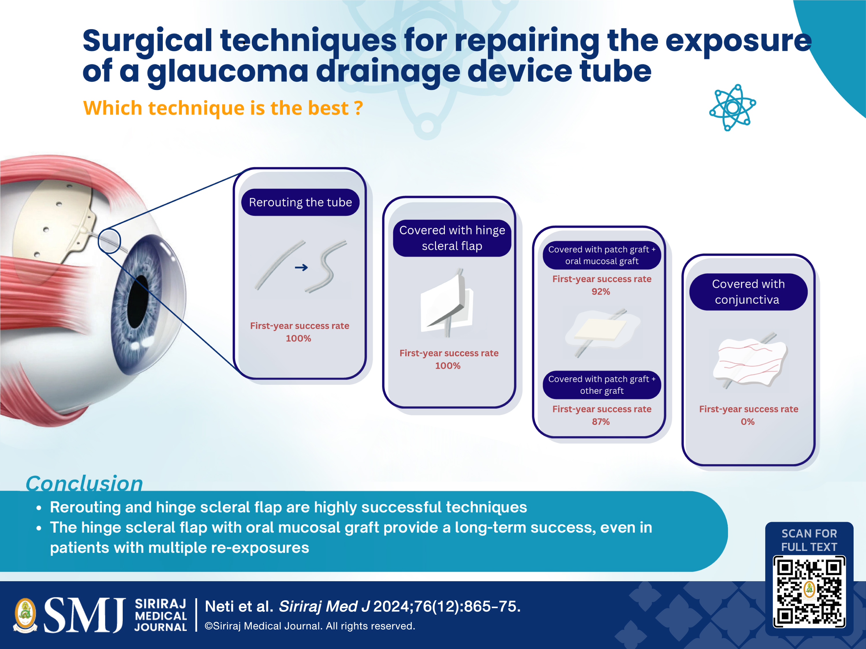
Results: We reported nine cases of GDD tube exposure repair, with additional 109 cases from 24 previous studies. One of our challenging cases encountered multiple tube revision failures by the patch graft technique; the exposure issue was sustainably resolved by a hinge scleral flap with buccal mucosal graft technique. Of the 118 cases, various surgical techniques were used, including patch grafts, hinge scleral flaps, primary conjunctival closure and rerouting. Among the cases, 61.6% were classified as difficult cases. The overall first, fifth and thirteenth-year survival rate
was 90.7%, 86.2% and 86.2%, respectively. Rerouting and scleral flap/tunnel techniques demonstrated the highest survival rate. No statistically significant differences in survival outcomes were observed among patch graft, scleral flap/tunnel and rerouting method (P = 0.129). The mean survival duration was 33.54 months. The duration of follow-up was 35.01 months.
Conclusion: Surgical management of GDD tube exposure yields favorable outcomes. A hinge scleral flap with buccal mucosal grafts can be a good option to treat challenging cases. The findings can shape an algorithm to manage GDD tube exposure.
References
Petchyim S, Subhadhirasakul A, Sakiyalak D, Vessadapan P, Ruangvaravate N. Clinical Characteristics and Outcome of Bleb-Related Infection in Glaucoma Patients. Siriraj Med J. 2022;74:555-561.
Rosentreter A, Lappas A, Widder RA, Alnawaiseh M, Dietlein TS. Conjunctival repair after glaucoma drainage device exposure using collagen-glycosaminoglycane matrices. BMC Ophthalmol. 2018;18(1):60.
Merrill KD, Suhr AW, Lim MC. Long-term success in the correction of exposed 5glaucoma drainage tubes with a tube extender. Am J Ophthalmol. 2007;144:136-7.
Joos KM, Laviña AM, Tawansy KA, Agarwal A. Posterior repositioning of glaucoma implants for anterior segment complications. Ophthalmology. 2001;108:279-84.
Prasher P, Lehmann JD, Aggarwal NK. Ahmed tube exposure secondary to prokera implantation. Eye Contact Lens. 2008;34(4):244-5.
Chun YS, Kim KW, Kim JC. Autologous tragal perichondrium patch graft for ahmed glaucoma valve tube exposure. J Glaucoma. 2013;22:e27-30.
Rosentreter A, Schild AM, Dinslage S, Dietlein TS. Biodegradable implant for tissue repair after glaucoma drainage device surgery. J Glaucoma. 2012;21:76-78.
Song YJ, Kim S, Yoon GJ. Case series: Use of stromal lenticule as patch graft. Am J Ophthalmol Case Rep. 2018;12:79-82.
Singh M, Chew PT, Tan D. Corneal patch graft repair of exposed glaucoma drainage implants. Cornea. 2008;27:1171-3.
Choudhari NS, Neog A, Latka S, Srinivasan B. Fibrin sealant-assisted revision of the exposed Ahmed tube. Middle East Afr J Ophthalmol. 2015;22:115-6.
Grover DS, Merritt J, Godfrey DG, Fellman RL. Forniceal conjunctival pedicle flap for the treatment of complex glaucoma drainage device tube erosion. JAMA Ophthalmol. 2013;131(5):662-6.
Godfrey DG, Merritt JH, Fellman RL, Starita RJ. Interpolated conjunctival pedicle flaps for the treatment of exposed glaucoma drainage devices. Arch Ophthalmol. 2003;121:1772-5.
Guajardo JM, Lim KS. Long-term Safety and Efficacy of Conjunctival Pedicle Graft Revision Combined With Repeated Pericardium Allograft for Exposed Glaucoma Drainage Devices. J Glaucoma. 2018;27:910-3.
Mohan S, Khattri M, Sah K, Pandey J, Sachan SK. Management of Tube Exposure Following Ahmed Glaucoma Valve Implantation by Allograft Corneoscleral Rim Patch. J Glaucoma. 2019;28:e67-e68.
Berezina TL, Fechtner RD, Cohen A, Kim EE, Chu DS. New Technique of Exposed Glaucoma Drainage Tube Repair: Report of a Case. J Curr Glaucoma Pract. 2015;9:62-64.
Ainsworth G, Rotchford A, Dua HS, King AJ. A novel use of amniotic membrane in the management of tube exposure following glaucoma tube shunt surgery. Br J Ophthalmol. 2006;90:417-9.
Jabbour S, Lesk MR, Harissi-Dagher M. Patch graft using collagen matrix (Ologen) for glaucoma drainage device exposure in a patient with Boston Keratoprosthesis type 1. Am J Ophthalmol Case Rep. 2018;12:32-35.
Mansoori T. Recurrent scleral patch graft shrinkage and Ahmed valve tube exposure. Nepal J Ophthalmol. 2019;11:232-6.
Einan-Lifshitz A, Belkin A, Mathew D, Sorkin N, Chan CC, Buys YM, et al. Repair of Exposed Ahmed Glaucoma Valve Tubes: Long-term Outcomes. J Glaucoma. 2018;27:532-6.
Alvarez-Ascencio D, Lazcano-Gomez G, Flores-Reyes E, Dueñas-Angeles K, Jímenez-Roman J, Kahook MY. Tenon Cyst Patch Graft for Ahmed Glaucoma Valve Tube Exposure: Case Series Report. J Glaucoma. 2021;30:e367-e71.
Lama PJ, Fechtner RD. Tube erosion following insertion of a glaucoma drainage device with a pericardial patch graft. Arch Ophthalmol. 1999;117:1243-44.
Liu X, Law SK. Autologous Partial-thickness Scleral Flap and Donor Corneal Graft in Management of Tube Erosion of Glaucoma Drainage Device. J Glaucoma. 2019;28:347-51.
Lee ES, Kang SY, Kim NR, Hong S, Ma KT, Seong GJ, et al. Split-thickness hinged scleral flap in the management of exposed tubing of a glaucoma drainage device. J Glaucoma. 2011;20:319-21.
Nardi M, Maglionico MN, Posarelli C, Figus M. Managing Ahmed Glaucoma Valve tube exposure: Surgical technique. Eur J Ophthalmol. 2021;31:778-81.
Page MJ, McKenzie JE, Bossuyt PM, Boutron I, Hoffmann TC, Mulrow CD, et al. The PRISMA 2020 statement: an updated guideline for reporting systematic reviews. BMJ. 2021;372:n71.
Murad MH, Sultan S, Haffar S, Bazerbachi F. Methodological quality and synthesis of case series and case reports. BMJ Evid Based Med. 2018;23:60-63.
Liu WW, Werner A, Chen TC. Repair of Tube Erosion by Modifying the Tube Extender. J Glaucoma. 2020;29:604-6.
Ehrlich HP, Hunt TK. Effects of cortisone and vitamin A on wound healing. Ann Surg. 1968;167:324-8.
Liang H, Baudouin C, Daull P, Garrigue JS, Brignole-Baudouin F. Ocular safety of cationic emulsion of cyclosporine in an in vitro corneal wound-healing model and an acute in vivo rabbit model. Mol Vis. 2012;18:2195-204.
Smith MF, Doyle JW, Ticrney JW, Jr. A comparison of glaucoma drainage implant tube coverage. J Glaucoma. 2002;11:143-7.
Silva N, Bollemeijer J, Ferreira A, Menéres M-J, Lemij H. Donor scleral graft vs pericardial graft vs scleral flap in tube drainage covering: advantages and disadvantages. Expert Review of Ophthalmology. January 2022; 1-10. Available from: Taylor & Francis Online. Accessed August 29, 2023.
Prabhasawat P. Amniotic membrane: a treatment for prevention of blindness from various ocular diseases. Siriraj Med J. 2007;59:139-41.
Mai C, Bertelmann E. Oral mucosal grafts: old technique in new light. Ophthalmic Res. 2013;50:91-98.
Published
How to Cite
License
Copyright (c) 2024 Siriraj Medical Journal

This work is licensed under a Creative Commons Attribution-NonCommercial-NoDerivatives 4.0 International License.
Authors who publish with this journal agree to the following conditions:
Copyright Transfer
In submitting a manuscript, the authors acknowledge that the work will become the copyrighted property of Siriraj Medical Journal upon publication.
License
Articles are licensed under a Creative Commons Attribution-NonCommercial-NoDerivatives 4.0 International License (CC BY-NC-ND 4.0). This license allows for the sharing of the work for non-commercial purposes with proper attribution to the authors and the journal. However, it does not permit modifications or the creation of derivative works.
Sharing and Access
Authors are encouraged to share their article on their personal or institutional websites and through other non-commercial platforms. Doing so can increase readership and citations.


