
Enhancing Orbital MRI Quality: Optimization of Saturation Pads for 1.5T
DOI:
https://doi.org/10.33192/smj.v77i5.271939Keywords:
Fat suppression, Orbit, MRI, Saturation pad, SPIRAbstract
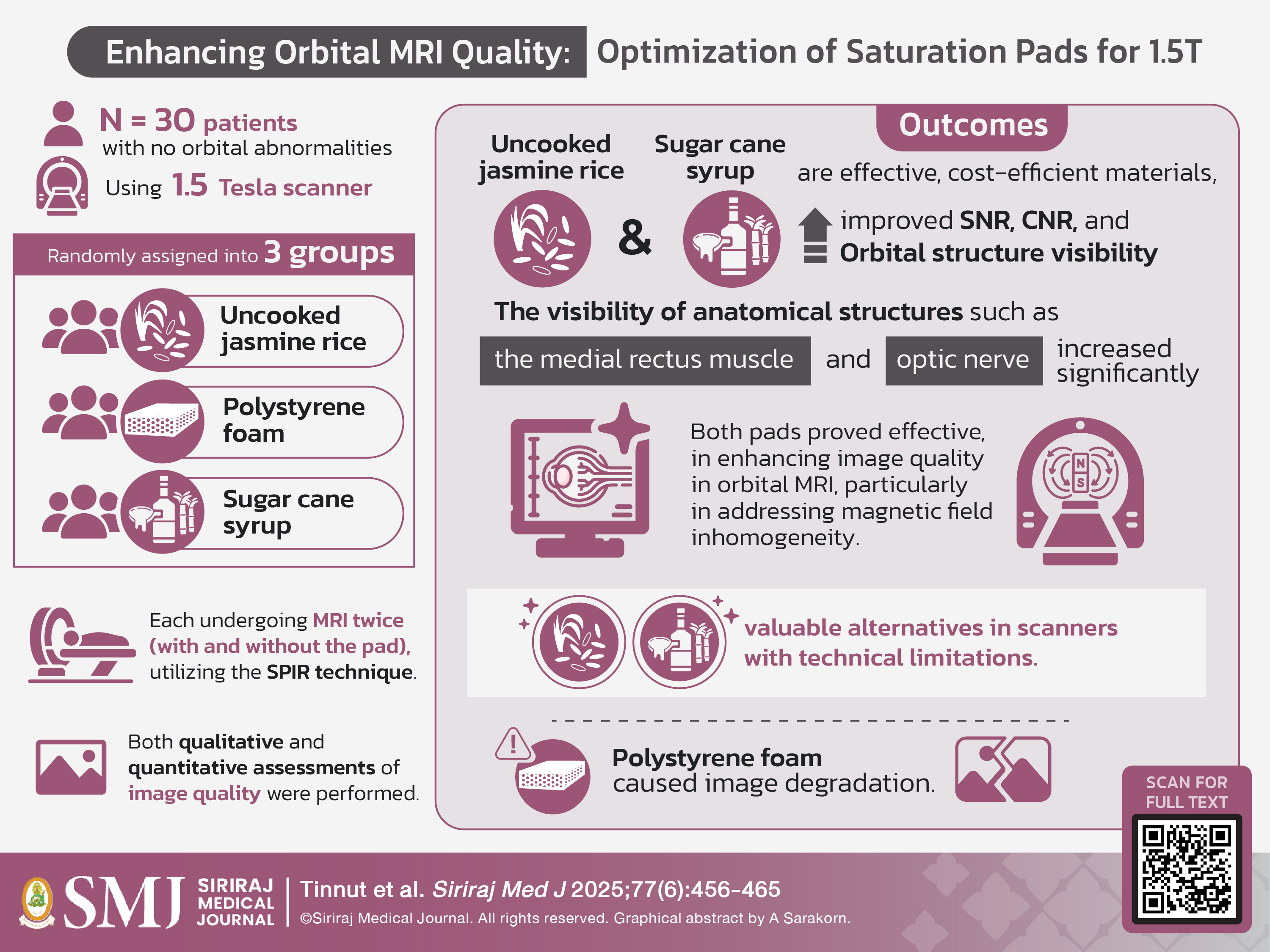
Objective: This study aimed to compare the effectiveness of different eye pads in enhancing magnetic field homogeneity using the Spectral Presaturation with Inversion Recovery (SPIR) technique.
Materials and Methods: A prospective study was approved by the Ethics Committee and involved thirty patients undergoing orbital MRI with no current abnormalities. Patients were randomly assigned to one of three groups: uncooked jasmine rice, polystyrene ball bullet foam, or sugar cane syrup. Each patient underwent imaging twice - first without a pad and then with a pad on the eyelid. An experienced neuroradiologist, blinded to pad compositions, evaluated the images quantitatively and qualitatively. Statistical analyses included the Kruskal-Wallis rank test for signal-to-noise (SNR) and contrast-to-noise ratios (CNR), Dunn’s test for post-hoc comparisons, the Wilcoxon signed-rank test for qualitative pre- and post-pad differences, and Fisher’s exact test for group differences, with significance set at P < 0.05.
Results: The uncooked jasmine rice group showed higher SNR and CNR, particularly in the medial rectus muscle (MRM). Significant improvements in the visual scale were noted for the optic nerve sheath complex (P = 0.002), MRM (P = 0.005), motion artifact (P = 0.034), and susceptibility artifact (P = 0.030) in both the uncooked jasmine rice and sugar cane syrup groups. Notably, none of the participants in the rice group exhibited a degraded visual scale for MRM or increased susceptibility artifact after pad placement.
Conclusion: This study highlights the effectiveness of uncooked jasmine rice and sugar cane syrup as materials for enhancing orbital MRI quality, especially in earlier scanner models.
References
Kennedy TA, Corey AS, Policeni B, Agarwal V, Burns J, Harvey HB, et al. ACR Appropriateness Criteria® Orbits Vision and Visual Loss. J Am Coll Radiol [Internet]. 2018;15(5, Supplement):S116–31. Available from: https://www.sciencedirect.com/science/article/pii/S154614401830351X
Grech R, Cornish KS, Galvin PL, Grech S, Looby S, O’Hare A, et al. Imaging of adult ocular and orbital pathology--a pictorial review. J Radiol Case Rep. 2014;8(2):1–29.
Lee AG, Johnson MC, Policeni BA, Smoker WRK. Imaging for neuro-ophthalmic and orbital disease - a review. Clin Experiment Ophthalmol. 2009;37(1):30–53.
Simon J, Szumowski J, Totterman S, Kido D, Ekholm S, Wicks A, et al. Fat-suppression MR imaging of the orbit. AJNR Am J Neuroradiol. 1988;9(5):961–8.
Tien RD. Fat-suppression MR imaging in neuroradiology: techniques and clinical application. AJR Am J Roentgenol. 1992;158(2):369–79.
Moriya S, Miki Y, Miyati T, Kanagaki M, Yokobayashi T. Flour pads: Devices to improve CHESS fat suppression. Magn Reson Med Sci. 2014;13(1):33–8.
Teshigawara M, Ogura A, Uchiyama N, Koganezawa T. The Optimal Pad Material for Fat Suppression of Breast MRI. Nihon Hoshasen Gijutsu Gakkai Zasshi. 2017;73(8):664-71.
Shiina R, Ogura A, Takeuchi T, Nakano H. Improvement of Fat Suppressing Effect for Finger and Neck MRI by Using Small Glass Beads. Nihon Hoshasen Gijutsu Gakkai Zasshi 2019;75(5):446-53.
Moriya S, Miki Y, Kamishima T, Kanagaki M, Yokobayashi T, Ishikawa M. Fat-suppressed MR images of both hands obtained using CHESS can be improved by rice pads. Eur J Radiol [Internet]. 2012;81(9):2318-22. Available from: http://dx.doi.org/10.1016/j.ejrad.2011.06.021
Moriya S, Miki Y, Yokobayashi T, Yamamoto A, Kanagaki M, Komori Y, et al. Rice and perfluorocarbon liquid pads: Comparison of fat suppression effects. Acta Radiol. 2010;51(5):534–8.
Moriya S, Miki Y, Yokobayashi T, Yamamoto A, Kanagaki M, Komori Y, et al. Improved CHESS imaging with the use of rice pads: Investigation in the neck, shoulder, and elbow. J Magn Reson Imaging. 2010;31(6):1504–7.
Moriya S, Miki Y, Yokobayashi T, Kanagaki M, Komori Y, Ishikawa M. Rice pads: Novel devices for homogeneous fat suppression in the knee. Acta Radiol. 2010;51(2):175–8.
Kang Y, Lee JW, Koh YH, Hur S, Kim SJ, Chai JW, et al. New MRI Grading System for the Cervical Canal Stenosis. Am J Roentgenol [Internet]. 2011 Jul 23 [cited 2020 Apr 29];197(1):W134–40. Available from: http://www.ajronline.org/doi/10.2214/AJR.10.5560
Cheol KB, Deok PH, Seok PK, Min PS, Gu SJ, Ok KJ, et al. A Study on the Usefulness of Polystyrene Pad for Fat Suppression Improvement. J Korean Soc MR Technol Vol. 27(2):31–6.
Eilenberg SS, Tartar M, Mattrey RF. Reducing magnetic susceptibility differences using liquid fluorocarbon pads (sat pad): Results with spectral presaturation of fat. Artif Cells Blood Substit Immobil Biotechnol. 1994;22(4):1477–83.
Mukherji SK, Tart RP, Fitzsimmons J, Belden C, McGorray S, Guy J, et al. Fat-suppressed MR of the orbit and cavernous sinus: comparison of fast spin-echo and conventional spin-echo. Am J Neuroradiol [Internet]. 1994;15(9):1707 LP – 1714. Available from: http://www.ajnr.org/content/15/9/1707.abstract
Moriya S, Miki Y, Yokobayashi T, Kanagaki M, Komori Y, Fujimoto K, et al. Rice pads reduce geometric distortion of echo-planar diffusion-weighted images of the cervical spinal cord. Magn Reson Med Sci. 2011;10(1):65–9.
Jin X, Zhao Y, Richardson A, Moore L, Williams PS, Zborowski M, et al. Differences in magnetically induced motion of diamagnetic, paramagnetic, and superparamagnetic microparticles detected by cell tracking velocimetry. Analyst. 2008;133(12):1767–75.
Mehrmohammadi M, Oh J, Mallidi S, Emelianov SY. Pulsed magneto-motive ultrasound imaging using ultrasmall magnetic nanoprobes. Mol Imaging. 2011;10(2):102–10.
Ikeguchi H, Shonai T, Mikami A, Yazawa N, Takahashi T, Yamada K. [Evaluation of prototype for additional pad packing (polystyrene ball bullet) for homogenously fat suppressed magnetic resonance imaging]. Nihon Hoshasen Gijutsu Gakkai Zasshi. 2013;69(1):71–9.
Oilmungmool N, Chattrapiban T, Tinnut S, Galassi W. Age-Related Changes in Signal Intensity Ratio of Normal Clivus Bone Marrow on Magnetic Resonance Imaging. Siriraj Med J. 2022;74:381–7.
Published
How to Cite
License
Copyright (c) 2025 Siriraj Medical Journal

This work is licensed under a Creative Commons Attribution-NonCommercial-NoDerivatives 4.0 International License.
Authors who publish with this journal agree to the following conditions:
Copyright Transfer
In submitting a manuscript, the authors acknowledge that the work will become the copyrighted property of Siriraj Medical Journal upon publication.
License
Articles are licensed under a Creative Commons Attribution-NonCommercial-NoDerivatives 4.0 International License (CC BY-NC-ND 4.0). This license allows for the sharing of the work for non-commercial purposes with proper attribution to the authors and the journal. However, it does not permit modifications or the creation of derivative works.
Sharing and Access
Authors are encouraged to share their article on their personal or institutional websites and through other non-commercial platforms. Doing so can increase readership and citations.


