
Sexual Dimorphism in Cranial and Post-Cranial Skeletal Elements: Forensic Implications for Sex Estimation in a Contemporary Thai Population
DOI:
https://doi.org/10.33192/smj.v77i11.277229Keywords:
Sexual dimorphism, cranial and post-cranial skeletal elements, forensic implications for sex estimation, contemporary Thai populationAbstract
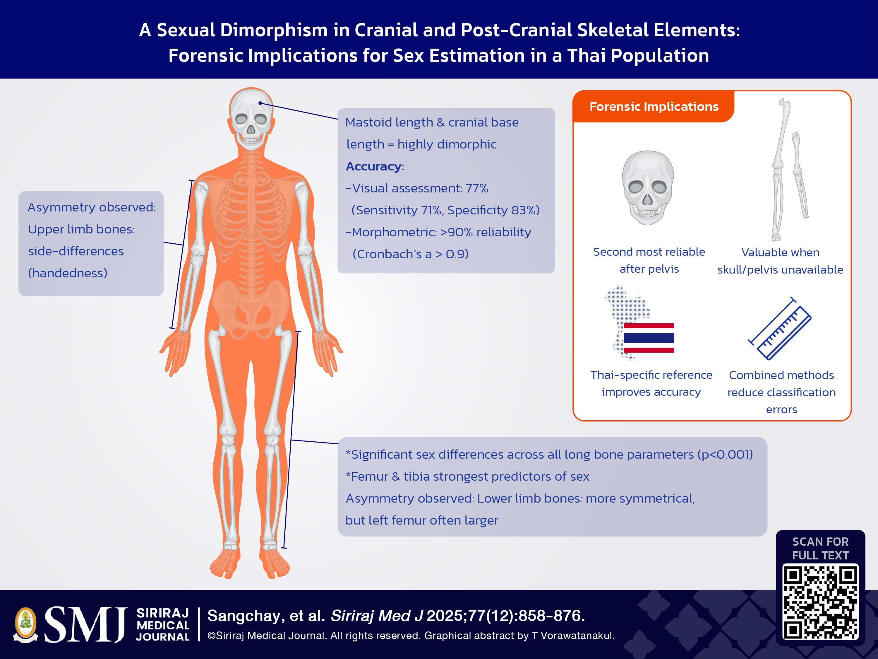
Objective: The analysis of human skeletal remains is instrumental in forensic and anthropological contexts, particularly for establishing biological profiles of unidentified individuals. Determining sex via skeletal examination is a fundamental component of this process and traditionally involves morphological assessment and metric analysis of pelvic and cranial bones. Nevertheless, the precision and reliability of these methodologies—whether through morphological evaluation or morphometric analysis—remain subjects of ongoing debate and scrutiny. This research set forth to investigate the efficacy of morphological and morphometric analysis in sex estimation by focusing on cranial and post-cranial long bones within contemporary Thai population.
Materials and Methods: The study sample comprised 204 skulls (105 from males, and 99 from females) and 200 sets of long bones of upper (humerus, radius, and ulna) and lower extremities (femur and tibia). Multiple measurements were systematically obtained from various anatomical regions of each bone, and measurements of extremity long bones were compared between the left and right sides.
Results: The analysis revealed statistically significant differences in these metrics between sexes, indicating the potential utility of this approach for sex classification. However, despite achieving high levels of accuracy, the studied methodology yielded some classification errors, which suggests some potential limitations.
Conclusion: The findings suggest that both inherent skeletal variability in cranial and post-cranial morphology within contemporary Thai population and the specific analytical techniques employed can markedly influence the accuracy of sex determination. These factors harbor and confer important implications for forensic and anthropological applications.
References
Wang X, Liu G, Wu Q, Zheng Y, Song F, Li Y. Sex estimation techniques based on skulls in forensic anthropology: a scoping review. Egypt J Forensic Sci. 2024;19(12):e0311762.
Lye R, Min H, Dowling J, Obertová Z, Estai M, Bachtiar NA, et al. Deep learning versus human assessors: forensic sex estimation from three-dimensional computed tomography scans. Sci Rep. 2024;14:30136.
Duangto P, Mahakkanukrauh P. Sex estimation from upper limb bones in a Thai population. Anat Cell Biol. 2020;53(1):36–43.
Krishan K, Chatterjee PM, Kanchan T, Kaur S, Baryah N, Singh RK. A review of sex estimation techniques during examination of skeletal remains in forensic anthropology casework. Forensic Sci Int. 2016;261:165.e1–8.
Selliah P, Martino F, Cummaudo M, Indra L, Biehler-Gomez L, Campobasso CP, et al. Sex estimation in middle and late adulthood: reliability of pelvic morphological traits and long bone metrics on an Italian skeletal collection. Forensic Sci Int. 2020; 134(5):1683-690
Blanc M, Knecht S, Nguyen K, Poulain C, Quatrehomme G, Alunni V, et al. Sexual dimorphism of the humerus bones in a French sample: comparison of several statistical models including machine learning models. Int J Legal Med. 2025;139(3):1395–408.
Houston SK, Brits D, Myburgh J, Liebenberg L. The impact of age-related changes in the skull on sex estimation using morphoscopic traits. Int J Legal Med. 2025.
Lewis CJ, Garvin HM. Reliability of the Walker cranial nonmetric method and implications for sex estimation. J Forensic Sci. 2016;61(3):743-51.
Wysocka J, Cieslik A, Danel D. Sex estimation using measurements of the proximal femur in a historical population from Poland. Anthropological Review. 2023;86(1):37-49.
Fliss B, Lüthi M, Fürnstahl P, Christensen AM, Sibold K, Thali M, et al. CT-based sex estimation on human femora using statistical shape modeling. Am J Phys Anthropol. 2019;169(2):279–86.
Setiawati R, Rahardjo P, Ruriana I, Guglielmi G. Anthropometric study using three-dimension pelvic CT scan in sex determination among adult Indonesian population. Forensic Sci Med Pathol. 2022;19(1):24-33.
Torimitsu S, Makino Y, Saitoh H, SakumaA, Ishii N, Yajima D, et al. Morphometric analysis of sex differences in contemporary Japanese pelves using multidetector computed tomography. Forensic Sci Int. 2015;257:530.e.1-7.
Houston SK, Brits D, Myburgh J, Liebenberg L. The impact of age-related changes in the skull on sex estimation using morphoscopic traits. Int J Legal Med. 2025.
Tallman SD. Cranial nonmetric sexual dimorphism and sex estimation in East and Southeast Asian individuals. Forensic Anthropology. 2019;2(4):204–21.
Lye R, Obertová Z, Bachtiar NA, Franklin D. Validating the use of clinical MSCT scans for cranial nonmetric sex estimation in a contemporary Indonesian population. Int J Legal Med. 2024;138(4):1559–71.
Pérez-Criado L, Rosas A, Bastir M, Pastor JF. Humeral laterality in modern humans and Neanderthals: a 3D geometric morphometric analysis. Anthropol Sci. 2017;125(3):117–28.
Kumar S, Voracek M, Singh M. The effects of hand preference and sex on right–left asymmetry in dorsal digit lengths among adults and children. Early Hum Dev. 2021;153:105293.
Langley NR, Jantz LM, McNulty S, Maijanen H, Ousley SD, Jantz RL. Error quantification of osteometric data in forensic anthropology. Forensic Sci Int. 2018;287:183-9.
Sangchay, N, Dzetkuličová, V, Zuppello M, Chetsawang J. Consideration of Accuracy and Observational Error Analysis in Pelvic Sex Assessment: A Study in a Thai Cadaveric Human Population. Siriraj Med J. 2022;74(5):330–9.
Rogers TL. Sex determination of human remains through cranial morphology. J Forensic Sci. 2005;50(3):493-500.
Kruger GC, L’Abbe’ EN, Stull KE, Kenyhercz MW. Sexual dimorphism in cranial morphology among modern South Africans. Int J Legal Med. 2015;129(4):869–75.
Mahakkanukrauh P, Sinthubua A, Prasitwattanaseree S, Ruengdit S, Singsuwan P, Praneatpolgrang S, et al. Craniometric study for sex determination in a Thai population. Anat Cell Biol. 2015;48(4):275–83.
Sinthubua A, Ruengdit S, Das S, Mahakkanukrauh P. A new method for sex estimation from maxillary suture length in a Thai population. Anat Cell Biol. 2017;50(4):261–4.
Saini V, Srivastava R, Rai RK, Shamal SN, Singh TB, Tripathi SK. Sex estimation from the mastoid process among North Indians. J Forensic Sci. 2012;57(2):434-9.
Ekizoglu O, Hocaoglu E, Inci E, Can IO, Solmaz D, Aksoy S, et al. Assessment of sex in a modern Turkish population using cranial anthropometric parameters. Leg Med (Tokyo). 2016;21:45-52.
Monum T, Prasitwattanaseree S, Das S, Siriphimolwat P, Mahakkanukrauh P. Sex estimation by femur in modern Thai population. Clin Ter. 2017;168(3):e203–e207.
Duangto P, Mahakkanukrauh P. Sex estimation from upper limb bones in a Thai population. Anat Cell Biol. 2020;53(1):36–43.
Ozer I, Katayama K. Sex determination using the femur in an ancient Japanese population. Coll Antropol. 2008;32(1):67-72.
Ranaweera L, Cabral E, Dissanayake DMPV, Lakshan WSV. Estimation of sex from the osteometric measurements of the femur in a contemporary Sri Lankan population. Int J Morphol. 2022;40(4):1009–17.
Cho H-J, Kwak D-S, Kim I-B. Morphometric evaluation of Korean femurs by geometric computation: comparisons of the sex and the population. BioMed Res Int. 2015;2015:730538.
Kim J-B, Lyu S-J, Kang H-W. Are Western knee designs dimensionally correct for Korean women? A morphometric study of resected femoral surfaces during primary total knee arthroplasty. Clin Orthop Surg. 2016;8(3):254–61.
Nanayakkara D, Vadysinghe AN, Nawarathna LS, Sampath H. Determination of sex from the tibia in a contemporary Sri Lankan population. J Forensic Sci Med. 2019;5(1):24–28.
Uehara K, Kadoya Y, Kobayashi A, Yamano Y. Anthropometry of the proximal tibia to design a total knee prosthesis for the Japanese population. J Arthroplasty. 2002;17(8):1028–32.
Özer BK, Özer İ, Sağır M, Güleç E. Sex determination using the tibia in an ancient Anatolian population. Mediterr Archaeol Archaeom. 2014;14(2):329–36.
Gupta C, Nayak N, Kalthur SG, D'Souza AS. A morphometric study of tibia and its nutrient foramen in South Indian population with its clinical implications. Saudi J Sports Med. 2015;15(3):244–8.
Nanayakkara D, Vadysinghe AN, Nawarathna LS, Sampath H. Determination of sex from the tibia in a contemporary Sri Lankan population. J Forensic Sci Med. 2019;5(1):24–28.
Tiwari A, Mahendru A, Priya A. An anatomical study of the tibia in the North Indian population. Int J Hum Anat. 2019;2(1):1–7.
Misiani MK, Amuti T, Darbar S, Mandela P, Maranga E, Obimbo M. Sex determination from dimensions of distal tibiae in adult Kenyans: A discriminant function analysis. Translational Research in Anatomy. 2020;20:100075.
Torres HR, Morais P, Fritze A, Burkhardt W, Kaufmann M, Oliveira B, et al. Anthropometric landmarking for diagnosis of cranial deformities: Validation of an automatic approach and comparison with intra- and interobserver variability. Ann Biomed Eng. 2022;50(9):1022–37.
Kotěrová AP, Santos F, Bejdová S, Rmoutilová R, Attia MH, Habiba A, et al. Prioritizing a high posterior probability threshold leading to low error rate over high classification accuracy: the validity of MorphoPASSE software for cranial morphological sex estimation in a contemporary population. Int J Legal Med. 2024;138(2):1759–68.
Arigbabu OA, Liao IY, Abdullah N, Mohamad Noor MH. Computer vision methods for cranial sex estimation. IPSJ Trans Comput Vis Appl. 2017;9:19.
Koo TK, Li MY. A guideline of selecting and reporting intraclass correlation coefficients for reliability research. J Chiropr Med. 2016;15(2):155–63.
Richard AH, Parks CL, Monson KL. Accuracy of standard craniometric measurements using multiple data formats. Forensic Sci Int. 2014;242:177–85.
Kizilgoz V, Aydin S, Aydemir H, Keles P, Kantarci M. Interobserver and intraobserver reliability of skull base angles measured on magnetic resonance images. World J Clin Cases. 2024;12(34):6687–95.
Walters J, Koo WWK, Bush A, Hammami M. Effect of hand dominance on bone mass measurement in sedentary individuals. J Clin Densitom. 1998;1(4):359–67.
Nandi ME, Olabiyi O, Okubike EA, Cyprain IE. A study of bilateral asymmetry of upper extremity and its effects on stature reconstruction amongst Nigerians. Aus J Forensic Sci. 2018;1(8):978–88.
Auerbach BM, Ruff CB. Limb bone bilateral asymmetry: variability and commonality among modern humans. J Hum Evol. 2006;50(2):203–18.
Marques S, Pinto C, Ferreira MT, Garcia S, Curate F. Sex Estimation from the Fibula and Tibia: A Study in Three Portuguese Reference Collections. Forensic Sci. 2023;5(1):2.
Mittino G, Langstaff H, García-Donas JG. Sex and Stature Estimation on the Tibia: A Virtual Pilot Study on a Contemporary Hispanic Population. J R Anthropol Inst. 2024;30(3):1–15.
Carvallo D, Retamal R. Sex estimation using the proximal end of the femur on a modern Chilean sample. Forensic Sci Int Rep. 2020;2:100077.
van der Gaast N, Dunning H, Huitema JM, Waters A, Jaarsma RL, Doornberg JN, et al. The symmetry of the left and right tibial plateau: a comparison of 200 tibial plateaus. Eur J Trauma Emerg Surg. 2023;49(1):69–74.
Verbakel J, Boot MR, van der Gaast N, Dunning H, Bakker M, Jaarsma RL, et al. Symmetry of the left and right tibial plafond; a comparison of 75 distal tibia pairs. Eur J Trauma Emerg Surg. 2024;50(6):2877–82.
Published
How to Cite
License
Copyright (c) 2025 Siriraj Medical Journal

This work is licensed under a Creative Commons Attribution-NonCommercial-NoDerivatives 4.0 International License.
Authors who publish with this journal agree to the following conditions:
Copyright Transfer
In submitting a manuscript, the authors acknowledge that the work will become the copyrighted property of Siriraj Medical Journal upon publication.
License
Articles are licensed under a Creative Commons Attribution-NonCommercial-NoDerivatives 4.0 International License (CC BY-NC-ND 4.0). This license allows for the sharing of the work for non-commercial purposes with proper attribution to the authors and the journal. However, it does not permit modifications or the creation of derivative works.
Sharing and Access
Authors are encouraged to share their article on their personal or institutional websites and through other non-commercial platforms. Doing so can increase readership and citations.


