
Calcaneal Articular Talar Facets, Stieda’s Process, and Calcaneus Secundarius: Variations Found in Thai Population
DOI:
https://doi.org/10.33192/smj.v77i12.277342Keywords:
Calcaneus, Talus, Facet, Prevalence, Anatomy, MorphologyAbstract
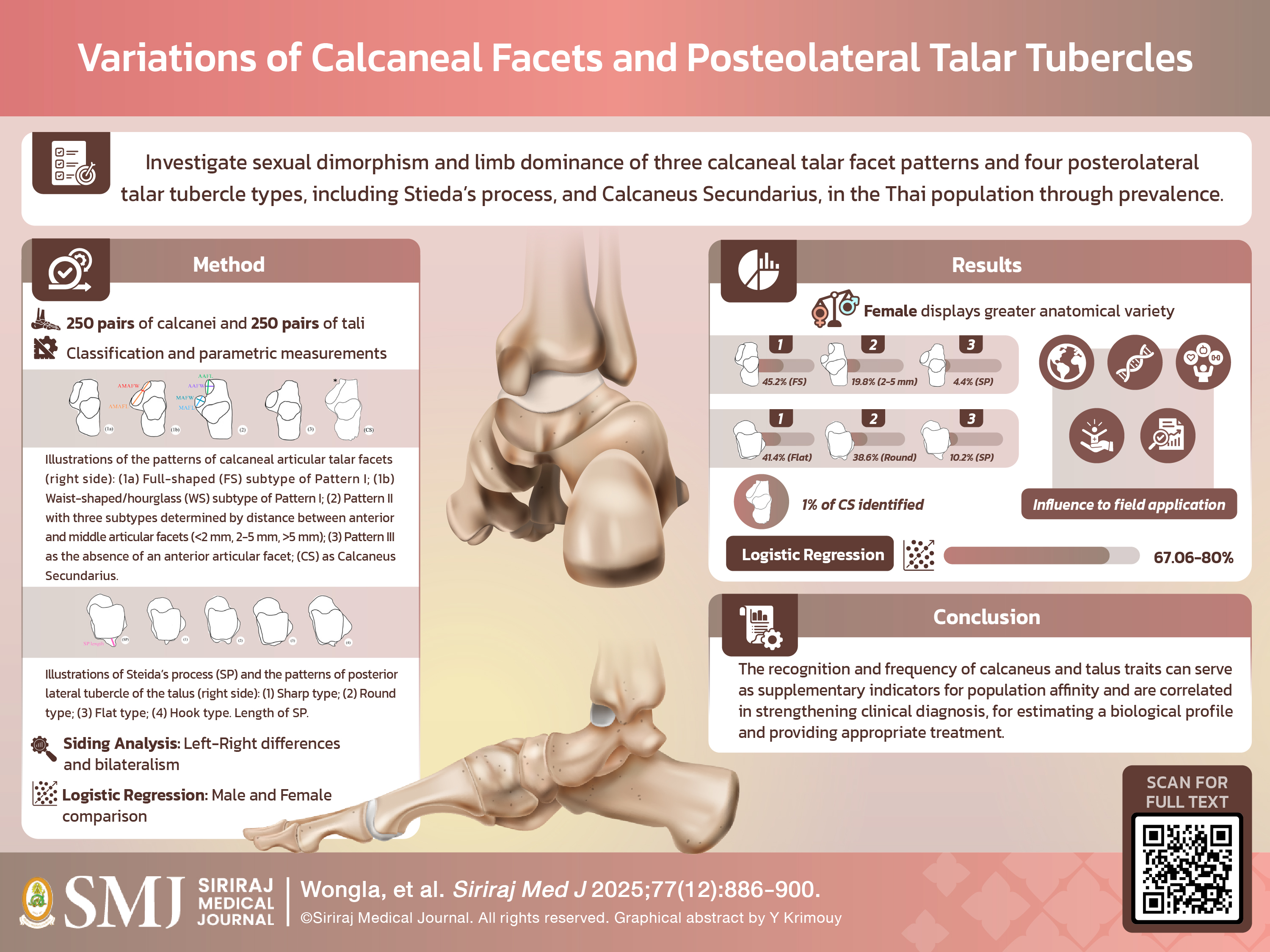
Objective: The calcaneus and talus have been studied in anatomy and pathology, but research is limited compared to its forensic use. This study examines the prevalence of three types of variants (calcaneal talar facet types, Stieda’s process, and calcaneus secundarius) to find correlations with sex and siding of the calcaneus and talus in a Thai
population.
Materials and Methods: A total of 250 specimens from the bone collection of Siriraj Anatomical and Anthropological Bone Research Centre (Si-AABRC), Thailand, were used. The calcaneus was classified into three types based on its articular talar facets, and the talus was categorized based on the presence of Stieda’s process and four types of posterolateral tubercles. Presence of a calcaneus secundarius was checked for a crescent-shaped notch and an accessory ossicle.
Results: Pattern I, the most common facet type, accounted for 60.23% of calcanei, with a 45.2% average of “fullshaped” facets. Pattern III was the least common, averaging 4.4%. The flat posterior processes of the talus were the most frequent trait (41.4%), while the hook type was the least common (2.4%). Calcaneus secundarius (CS) and Stieda’s process (SP) were the rarest traits.
Conclusion: The aforementioned calcaneal and talar traits can aid in investigating sides, as left or right, but not as a specific sex characteristic. The calcaneus secundarius has no correlation to either sex or siding due to limited samples. The outcomes of prevalence align with prior studies.
References
Hall RL, Shereff MJ. Anatomy of the calcaneus. Clinical Orthopaedics and Related Research (1976-2007). 1993;290:27–35.
Gupton M, Özdemir M, Terreberry RR. Anatomy, bony pelvis and lower limb: calcaneus. In: StatPearls [Internet]. Treasure Island (FL): StatPearls Publishing; 2025 Jan.
Cockerill SJ, Arnay‐de‐la‐Rosa M, González‐Reimers E. An atlas of anatomical variants of the human calcaneus. J Morphol. 2024;285(5):e21706.
Artkengkla T, Piyawinijwong S. Variations of Calcaneal Articular Facets in Thais Related to Navicular Facet ความแตกต่างของ ชนิดข้อต่อบนกระดูกแคลคาเนียส และความเกี่ยวข้องกับข้อ ต่อบนกระดูกนาวิคูลาร์ในคนไทย. 34th National Graduate Research Conference. 2015.
Angelova M, Marinova D. Anatomical Variations of the Articular Surfaces of the Calcaneus among Bulgarian Population. Acta Morphologica et Anthropologica. 2024;31:1–2.
Prasad SA, Rajasekhar S. Morphometric analysis of talus and calcaneus. Surg Radiol Anat. 2019;41(1):9–24.
Hegazy AAM, Hegazy MA. Talus bone: Unique anatomy. International Journal of Cadaveric Studies and Anatomical Variations. 2022;3(2):52–5.
Khan IA, Varacallo MA. Anatomy, bony pelvis and lower limb, foot talus. In: StatPearls [Internet]. Treasure Island (FL): StatPearls Publishing; 2025 Jan.
Boyan N, Ozsahin E, Kızılkanat E, Soames R, OĞUZ Ö. Morphometric measurement and types of articular facets on the talus and calcaneus in an Anatolian population. International Journal of Morphology. 2016;34(4).
Srivastava S, Khan AZ, Chigurupalli K, Singh KV, Arora NK, Haque M. Comparative Analysis of Squatting Facets on Femur, Tibia, and Talus: Insights from A Population in Northwest Uttar Pradesh and Cross–Population Comparisons. International Journal of Pharmaceutical and Clinical Research. 2023;15(6):87–96.
El-Eishi H. Variations in the talar articular facets in Egyptian calcanei. Cells Tissues Organs. 1974;89(1):134–8.
Yang Y, Cheng H-W, Xiong Z-R, Liu N, Liu Y, Wang Y, et al. Classification and morphological parameters of the calcaneal Talar facet: which type is more likely to cause osteoarthritis in Chinese population? Biomed Res Int. 2019;2019:6095315.
Lawrence RC, Hochberg MC, Kelsey JL, McDuffie FC, Medsger Jr TA, Felts WR, et al. Estimates of the prevalence of selected arthritic and musculoskeletal diseases in the United States. J Rheumatol. 1989;16(4):427–41.
Boonruangsri P, Woraputtaporn W, Namking M. The pattern of talar articular facets in Northeastern Thai calcanei. Srinagarind Hosp Med J. 1992;7(1):28–34.
Bidmos M. Metrical and non-metrical assessment of population affinity from the calcaneus. Forensic Sci Int. 2006;159(1):6–13.
Nozaki S, Watanabe K, Kamiya T, Katayose M, Ogihara N. Sex-and age-related morphological variations in the talar articular surfaces of the calcaneus. Ann Anat. 2020;229:151468.
Garg R, Dagal N, Kumar S, Shekhawat S. Study of patterns of talar articular facets of human calcanei and their clinical implications in population of Rajasthan. Indian J Basic Appl Med Res. 2013;7(2):643–50.
Vučinić N, Teofilovski-Parapid G, Erić M, Tubbs RS, Radošević D, Jovančević B. Morphometric analysis of the patterns of calcaneal facets for the talus in Serbian population. PLoS One. 2020;15(10):e0240818.
Spradley MK. Metric Methods for the Biological Profile in Forensic Anthropology: Sex, Ancestry, and Stature. Acad Forensic Pathol. 2016;6(3):391–9.
Singh DV. Assessment of morphometric study of calcaneus and its articular facets. Int J Acad Med Pharm. 2023;5(1):801–3.
Kumar S, Singh AK, Fatima N, Akhtar J, Ratnesh R, Kumar V. A morphological study on patterns of human calcaneal articular facets for talus in population of bihar and its clinical implications. Journal of Evolution of Medical and Dental Sciences. 2017;6(56):4193–7.
White TD, Black MT, Folkens PA. Human osteology: Academic press; 2011.
Mann RW, Hunt DR, Lozanoff S. Photographic regional atlas of non-metric traits and anatomical variants in the human skeleton: Charles C Thomas Publisher; 2016.
Mann RW. The bone book: A photographic lab manual for identifying and siding human bones: Charles C Thomas Publisher; 2017.
Mann R. Calcaneus secundarius. Variation of a common accessory ossicle. J Am Podiatr Med Assoc. 1989;79(8):363–6.
Silva AM, Curate F. Accessory foot bones in a Portuguese identified skeletal collection. Sci Rep. 2024;14(1):17169.
Ouamthong R, Mahacharoen T, Inthasan C, Srisinghasongkram J, Singsuwan P, Mahakkanukrauh P. A Comparison of Effectiveness of Sex Estimation from the Calcaneus and Talus in a Thai Population. Int J Morphol. 2023;41(1):268-77.
Scott S, Ruengdit S, Peckmann TR, Mahakkanukrauh P. Sex estimation from measurements of the calcaneus: Applications for personal identification in Thailand. Forensic Sci Int. 2017;278:405.e1–e8.
Derin Cicek E, Bankaoglu M. Prevalence of Elongated Posterior Talar Process (Stieda Process) Detected by Radiography. International Journal of Morphology. 2020;38(4).
Koshy S, Vettivel S, Selvaraj K. Estimation of length of calcaneum and talus from their bony markers. Forensic Sci Int. 2002;129(3):200–4.
Agarwal S, Garg S, Vasudeva N. Subtalar joint instability and calcaneal spurs associated with the configuration of the articular facets of adult human calcaneum in Indian population. J Clin Diagn Res. 2016;10(9):AC05-AC09.
Mann RW, Ubelaker DH. The forensic anthropologist. FBI L Enforcement Bull. 1990;59:20.
Kalbouneh H, Alsalem M, Hani MB, Alhusamiah H, Momani Y, Massad T, et al. A Comprehensive Study of the Anatomical Variations of the Posterolateral Tubercle of Talus. International Journal of Morphology. 2021;39(3).
Yang H, Liao L, Xue F, Li Y, Hu G. Anatomical observation, classification, fracture and finite element analysis of the posterior process of the Asian adult talus. J Orthop Surg Res. 2022;17(1):444.
Keles-Celik N, Kose O, Sekerci R, Aytac G, Turan A, Güler F. Accessory ossicles of the foot and ankle: disorders and a review of the literature. Cureus. 2017;9(11).
Jamovi. The jamovi project (Version 2.5).
Koukiasa AE, Eliopoulos C, Manolis SK. Biometric sex estimation using the scapula and clavicle in a modern Greek population. Anthropol Anz. 2017;74(3):241-6.
Ricklan D, Tobias P. Unusually low sexual dimorphism of endocranial capacity in a Zulu cranial series. Am J Phys Anthropol. 1986;71(3):285–93.
Robin X, Turck N, Hainard A, Tiberti N, Lisacek F, Sanchez J-C, et al. pROC: an open-source package for R and S+ to analyze and compare ROC curves. BMC Bioinformatics. 2011;12:77.
Heinze G, Ploner M, Dunkler D, Southworth H, Heinze MG. Package ‘logistf’. 2025. Available from: https://cran.r-project.org/web/packages/logistf/logistf.pdf
Firth D. Bias reduction of maximum likelihood estimates. Biometrika. 1993;80(1):27–38.
McShane PA. The prevalence of absent anterior facet of the calcaneus and a suggested convention for the naming of the configuration of the superior facets of the calcaneus. Int J Anat Res. 2021;9(1.3):7928–34.
Uygur M, Atamaz F, Celik S, Pinar Y. The types of talar articular facets and morphometric measurements of the human calcaneus bone on Turkish race. Arch Orthop Trauma Surg. 2009;129(7):909–14.
Sharada R, Sneha K, Gupta C, Pai SR, Rairam G. Non-metrical study of the pattern of talar articular facets in south Indian dry calcanei. Surg Radiol Anat. 2012;34(6):487–91.
Shweta J, Rashvaita K, Krunal R, Meenakshi B. Patterns of talar articular facets on calcaneum and its clinical implication. Int J Anat Physiol. 2013;2(4):23–6.
Das S, Sinthubua A, Ruengdit S, Mahakkanukrauh P. Anomalous Calcaneus Secundarius: Anatomical and Clinical Considerations. International Medical Journal. 2018;25(5).
Rühli FJ, Solomon L, Henneberg M. High prevalence of tarsal coalitions and tarsal joint variants in a recent cadaver sample and its possible significance. Clin Anat. 2003;16(5):411–5.
Iamsaard S, Uabundit N, Boonruangsri P, Sawatpanich T, Hipkaeo W. Types of Facets on the Superior Articular Surface of Isan-Thai Dried Calcanei. Int J Morphol. 2015;33(4):1549–52.
Harper CM. Human calcaneal variation relative to subsistence strategy, activity level, and footwear. Front Earth Sci. 2023;11:1213374.
Mowtoshee NR, Naushaba H, Kishwara S, Kamal AM, Rahman M, Singha PS. Variations In Talar Articular Facets on Dry Adult Human Left Calcaneus. Bangladesh Journal of Anatomy. 2020;18(1):17–20.
Drayer-Verhagen F. Arthritis of the subtalar joint associated with sustentaculum tali facet configuration. J Anat. 1993;183(Pt 3):631-4.
Agarwal P, Agarwal V, Kumar A, Kumar S, Gupta R. A Study of Morphological Pattern of the Talar Articular Facets in Dry Human Calcanei and Its Clinical Implications. International Journal of Health Sciences. 2022;6(S4):12676–83.
Cheng KY, Smitaman E, Resnick DL. Developmental Talocalcaneal Coalitions and Associated Conditions. MRI Web Clinic; 2022. Available from: https://radsource.us/wp-content/uploads/2022/06/2206_Tarsal-Coalition-Resnick_FINAL.pdf
Bruckner J. Variations in the Human Subtalar Joint. J Orthop Sports Phys Ther. 1987;8(10):489–94.
Anbumani S, Sridharan R, Selvi A. An anatomical study of morphology and morphometric analysis of calcaneum and its talar articular surfaces. International Journal of Anatomy and Research. 2017;5(3.2):4223–9.
Harnroongroj T, Chuckpaiwong B, Angthong C, Nanakorn P, Sudjai N, Harnroongroj T. Displaced articular calcaneus fractures: classification and fracture scores: a preliminary study. J Med Assoc Thai. 2012;95(3):366-77.
Ohishi T, Fujita T, Nishida T, Asukai M, Suzuki D, Matsuyama Y. Tibial spastic varus foot caused by os calcaneus secundarius: A case report. Foot (Edinb). 2019;39:92–5.
Çorbacıoğlu ŞK, Aksel G. Receiver operating characteristic curve analysis in diagnostic accuracy studies: A guide to interpreting the area under the curve value. Turk J Emerg Med. 2023;23(4):195–8.
Ogut E. The Stieda process of the talus: the anatomical knowledge and clinical significance of an overlooked protrusion. Bulletin of the National Research Centre. 2022;46(1):280.
Brodsky AE, Khalil MA. Talar compression syndrome. Am J Sports Med. 1986;14(6):472–6.
Jilani LZ, Istiyak M, Siddiqui YS. Stieda Process as a Source of Posterior Ankle Pain: A Case Report with Its Structural and Clinical Implication. Journal of Foot and Ankle Surgery (Asia Pacific). 2024;11(3):147–51.
Zwiers R, Baltes TP, Opdam KT, Wiegerinck JI, van Dijk CN. Prevalence of os trigonum on CT imaging. Foot Ankle Int. 2018;39(3):338–42.
Mann RW. Calcaneus secundarius: description and frequency in six skeletal samples. Am J Phys Anthropol. 1990;81(1):17–25.
Candan B, Torun E, Dikici R. The prevalence of accessory ossicles, sesamoid bones, and biphalangism of the foot and ankle: a radiographic study. Foot Ankle Orthop. 2022;7(1):24730114211068792.
Ceroni D, De Coulon G, Spadola L, De Rosa V, Kaelin A. Calcaneus secundarius presenting as calcaneonavicular coalition: a case report. J Foot Ankle Surg. 2006;45(1):25–7.
Published
How to Cite
License
Copyright (c) 2025 Siriraj Medical Journal

This work is licensed under a Creative Commons Attribution-NonCommercial-NoDerivatives 4.0 International License.
Authors who publish with this journal agree to the following conditions:
Copyright Transfer
In submitting a manuscript, the authors acknowledge that the work will become the copyrighted property of Siriraj Medical Journal upon publication.
License
Articles are licensed under a Creative Commons Attribution-NonCommercial-NoDerivatives 4.0 International License (CC BY-NC-ND 4.0). This license allows for the sharing of the work for non-commercial purposes with proper attribution to the authors and the journal. However, it does not permit modifications or the creation of derivative works.
Sharing and Access
Authors are encouraged to share their article on their personal or institutional websites and through other non-commercial platforms. Doing so can increase readership and citations.


