
Intraoperative Cerebral Angiography in the Surgical Resection of Brain Arteriovenous Malformations
DOI:
https://doi.org/10.33192/smj.v78i5.277427Keywords:
AVM, cerebral arteriovenous malformation, intraoperative cerebral angiography, residual, surgical resectionAbstract
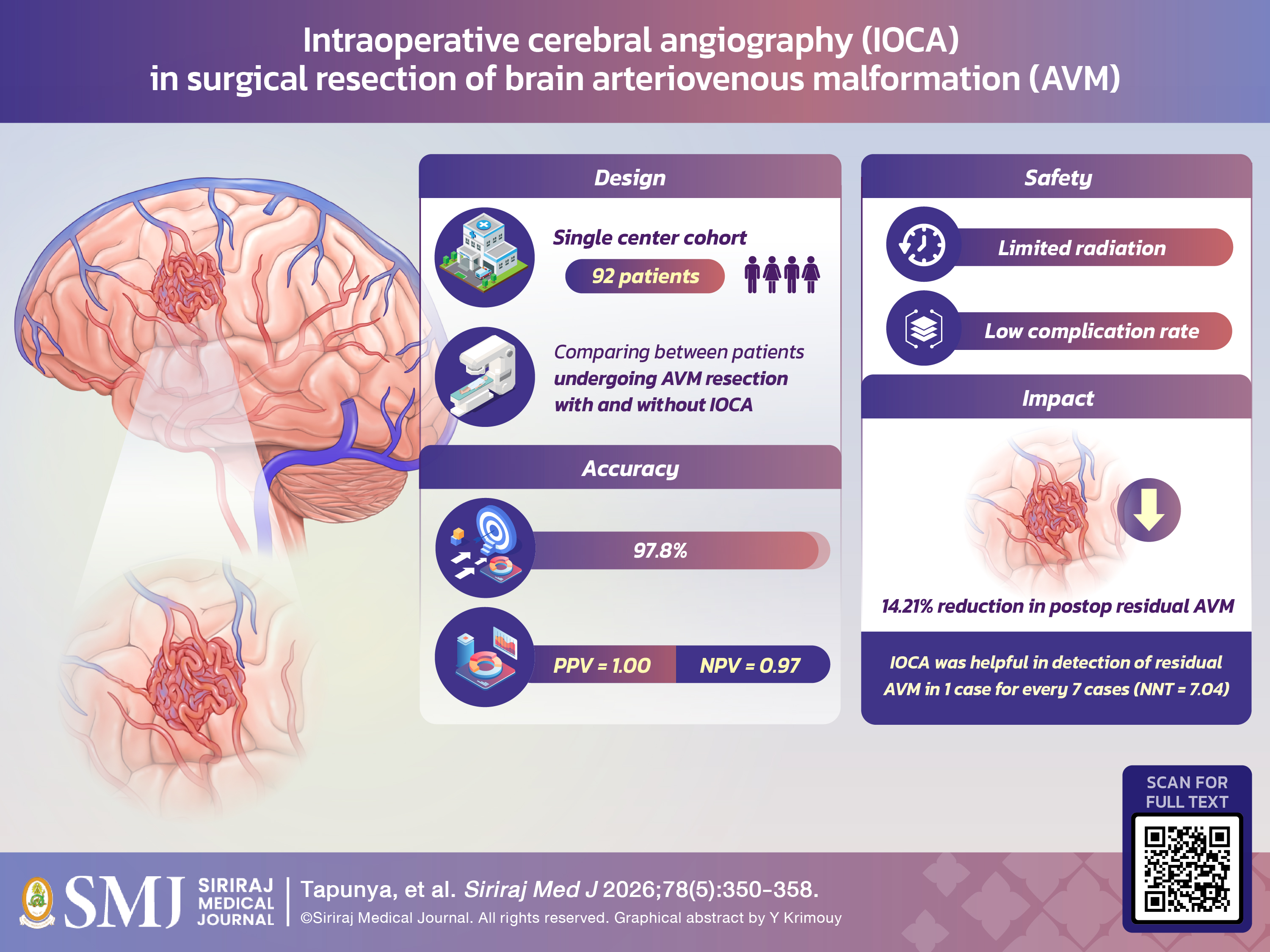
Objective: This study aimed to determine the effectiveness of IOCA in reducing residual AVMs after surgery.
Materials and Methods: We retrospectively reviewed all AVM resection surgeries performed at Siriraj Hospital between January 2008 and December 2022. The use of intraoperative cerebral angiography was recorded. The primary outcome was the presence of residual AVM on postoperative imaging
Results: The study included 92 patients, who were divided into two groups: those undergoing IOCA (44) and those undergoing surgery without IOCA (48). The initial incidence of residual AVMs prior to IOCA did not differ between the two groups (15.90% vs. 18.75%, p=0.720). However, postoperative imaging revealed significantly fewer missed residual AVMs in the IOCA group compared to the non-IOCA group (4.54% vs. 18.75%, p=0.036; absolute risk reduction (ARR) = 14.21% and a number needed to treat (NNT) = 7.04). The overall rate of acute complications did not differ significantly between the two groups (p = 0.108), but the mean length of hospital stay was higher in the non-IOCA group (19.31 vs. 8.57, p=0.001).
Conclusion: Intraoperative cerebral angiography may help reduce the incidence of residual AVMs after surgery and decrease the length of hospital stays.
References
Al-Shahi R, Bhattacharya JJ, Currie DG, Papanastassiou V, Ritchie V, Roberts RC, et al. Prospective, population-based detection of intracranial vascular malformations in adults: the Scottish Intracranial Vascular Malformation Study (SIVMS). Stroke. 2003;34(5):1163-9.
Al-Shahi R, Fang JS, Lewis SC, Warlow CP. Prevalence of adults with brain arteriovenous malformations: a community based study in Scotland using capture-recapture analysis. J Neurol Neurosurg Psychiatry. 2002;73(5):547-51.
ApSimon HT, Reef H, Phadke RV, Popovic EA. A population-based study of brain arteriovenous malformation: long-term treatment outcomes. Stroke. 2002;33(12):2794-800.
Fults D, Kelly DL, Jr. Natural history of arteriovenous malformations of the brain: a clinical study. Neurosurgery. 1984;15(5):658-62.
Madzar D, Kuramatsu JB, Gollwitzer S, Lucking H, Kloska SP, Hamer HM, et al. Seizures among long-term survivors of conservatively treated ICH patients: incidence, risk factors, and impact on functional outcome. Neurocrit Care. 2014;21(2):211-9.
Bilbao CJ, Bhalla T, Dalal S, Patel H, Dehdashti AR. Comparison of indocyanine green fluorescent angiography to digital subtraction angiography in brain arteriovenous malformation surgery. Acta Neurochir (Wien). 2015;157(3):351-9.
Bauer BL. Intraoperative angiography in cerebral aneurysm and AV-malformation. Neurosurg Rev. 1984;7(2-3):209-17.
Anegawa S, Hayashi T, Torigoe R, Harada K, Kihara S. Intraoperative angiography in the resection of arteriovenous malformations. J Neurosurg. 1994;80(1):73-8.
Chalouhi N, Theofanis T, Jabbour P, Dumont AS, Fernando Gonzalez L, Starke RM, et al. Safety and efficacy of intraoperative angiography in craniotomies for cerebral aneurysms and arteriovenous malformations: a review of 1,093 consecutive cases. Neurosurgery. 2012;71(6):1162-9.
Komatsu K, Takagi Y, Ishii A, Kikuchi T, Yamao Y, Yoshida K, et al. Changes in treatment strategy over time for arteriovenous malformation in a Japanese high-volume center. BMC Neurol. 2020;20(1):404.
Vitaz TW, Gaskill-Shipley M, Tomsick T, Tew JM, Jr. Utility, safety, and accuracy of intraoperative angiography in the surgical treatment of aneurysms and arteriovenous malformations. AJNR Am J Neuroradiol. 1999;20(8):1457-61.
Al-Shahi Salman R, White PM, Counsell CE, du Plessis J, van Beijnum J, Josephson CB, et al. Outcome after conservative management or intervention for unruptured brain arteriovenous malformations. JAMA. 2014;311(16):1661-9.
Spetzler RF, Martin NA. A proposed grading system for arteriovenous malformations. J Neurosurg. 1986;65(4):476-83.
Lawton MT, Kim H, McCulloch CE, Mikhak B, Young WL. A supplementary grading scale for selecting patients with brain arteriovenous malformations for surgery. Neurosurgery. 2010;66(4):702-13; discussion 13.
Hafez A, Koroknay-Pal P, Oulasvirta E, Elseoud AA, Lawton MT, Niemela M, et al. The Application of the Novel Grading Scale (Lawton-Young Grading System) to Predict the Outcome of Brain Arteriovenous Malformation. Neurosurgery. 2019;84(2):529-36.
Ihn YK, Kim BS, Byun JS, Suh SH, Won YD, Lee DH, et al. Patient Radiation Exposure During Diagnostic and Therapeutic Procedures for Intracranial Aneurysms: A Multicenter Study. Neurointervention. 2016;11(2):78-85.
Wonglao S. Evaluation of the Patient Radiation Doses from Computed Tomography (CT) in Neurological Institute of Thailand. Journal of the Department of Medical Services. 2022;47(3):51-9.
Lin EC. Radiation risk from medical imaging. Mayo Clin Proc. 2010;85(12):1142-6; quiz 6.
Gaballah M, Storm PB, Rabinowitz D, Ichord RN, Hurst RW, Krishnamurthy G, et al. Intraoperative cerebral angiography in arteriovenous malformation resection in children: a single institutional experience. J Neurosurg Pediatr. 2014;13(2):222-8.
Yoon W, Kim H, Kim YW, Kim SR, Park IS. Usefulness and Stability of Intraoperative Digital Subtraction Angiography Using the Transradial Route in Arteriovenous Malformation Surgery. World Neurosurg. 2018;111:e799-e805.
Ellis MJ, Kulkarni AV, Drake JM, Rutka JT, Armstrong D, Dirks PB. Intraoperative angiography during microsurgical removal of arteriovenous malformations in children. J Neurosurg Pediatr. 2010;6(5):435-43.
Gross BA, Frerichs KU, Du R. Sensitivity of CT angiography, T2-weighted MRI, and magnetic resonance angiography in detecting cerebral arteriovenous malformations and associated aneurysms. J Clin Neurosci. 2012;19(8):1093-5.
Lim HK, Choi CG, Kim SM, Kim JL, Lee DH, Kim SJ, et al. Detection of residual brain arteriovenous malformations after radiosurgery: diagnostic accuracy of contrast-enhanced four-dimensional MR angiography at 3.0 T. Br J Radiol. 2012;85(1016):1064-9.
Published
How to Cite
License
Copyright (c) 2026 Siriraj Medical Journal

This work is licensed under a Creative Commons Attribution-NonCommercial-NoDerivatives 4.0 International License.
Authors who publish with this journal agree to the following conditions:
Copyright Transfer
In submitting a manuscript, the authors acknowledge that the work will become the copyrighted property of Siriraj Medical Journal upon publication.
License
Articles are licensed under a Creative Commons Attribution-NonCommercial-NoDerivatives 4.0 International License (CC BY-NC-ND 4.0). This license allows for the sharing of the work for non-commercial purposes with proper attribution to the authors and the journal. However, it does not permit modifications or the creation of derivative works.
Sharing and Access
Authors are encouraged to share their article on their personal or institutional websites and through other non-commercial platforms. Doing so can increase readership and citations.


