
The Correlation between Chest X-ray and Cardiac Magnetic Resonance Imaging in the Assessment of Left Atrial Enlargement
DOI:
https://doi.org/10.33192/smj.v78i1.278423Keywords:
Left atrial enlargement, cardiac magnetic resonance imaging, chest X-rayAbstract
Objective: Left atrial enlargement (LAE) is common in cardiovascular disease and is associated with heart failure, atrial fibrillation, and stroke. Chest X-ray (CXR) is widely available; however, its diagnostic value for LAE has not been validated against cardiac magnetic resonance imaging (CMR). We evaluated the correlation and diagnostic performance of conventional CXR signs for detecting LAE using CMR as the reference standard.
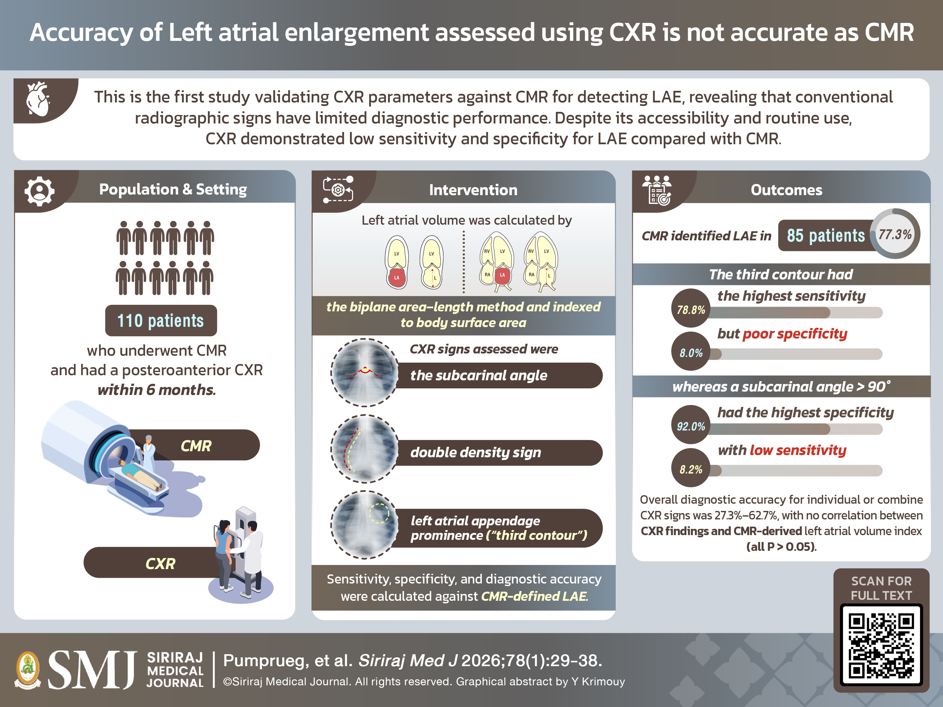
Materials and Methods: We retrospectively analyzed 110 patients who underwent CMR and had a posteroanterior CXR within 6 months. Left atrial volume was calculated by the biplane area–length method and indexed to body surface area. CXR signs assessed were the subcarinal angle, double density sign, and left atrial appendage prominence (“third contour”). Sensitivity, specificity, and diagnostic accuracy were calculated against CMR-defined LAE.
Results: CMR identified LAE in 85 patients (77.3%). The third contour had the highest sensitivity (78.8%) but poor specificity (8.0%), whereas a subcarinal angle > 90° had the highest specificity (92.0%) with low sensitivity (8.2%). Overall diagnostic accuracy for individual or combined CXR signs was 27.3%–62.7%, with no correlation between CXR findings and CMR-derived left atrial volume index (all P > 0.05).
Conclusions: To our knowledge, this is the first study validating CXR parameters against CMR for detecting LAE, revealing that conventional radiographic signs have limited diagnostic performance. Despite its accessibility and routine use, CXR demonstrated low sensitivity and specificity for LAE compared with CMR.
References
Haykal R, Kassar A, Chamoun N, Akoum N. The left atrium in heart failure with preserved ejection fraction: What we know and what we do not know. Heart Rhythm O2. 2025;6(7):1028–38.
Thomas L, Abhayaratna WP. Left Atrial Reverse Remodeling: Mechanisms, Evaluation, and Clinical Significance. JACC Cardiovasc Imaging. 2017;10(1):65–77.
Janwanishstaporn S, Boonyasirinant T. Correlation between aortic stiffness and left atrial volume index in hypertensive patients. Clin Exp Hypertens. 2016;38(2):160–5.
Jaturapisanukul S, Kaolawanich Y, Meechuen M, Boonyasirinant T. Correlation between Obesity and Left Atrial Enlargement in Patients Using Cardiac Magnetic Resonance. Siriraj Med J. 2025;77(2):130–6.
Higgins CB, Reinke RT, Jones NE, Broderick T. Left atrial dimension on the frontal thoracic radiograph: a method for assessing left atrial enlargement. AJR Am J Roentgenol. 1978;130(2):251–5.
Quinton SJ, Ker JA, Rheeder P, Deffur A. The reliability of chest radiographs in predicting left atrial enlargement. Cardiovasc J Afr. 2010;21(5):274–9.
El Mathari S, Hopman L, Bouchnaf C, Heidendael JF, Nederveen AJ, van Ooij P, et al. Clinical implications of different methods to assess left atrial remodeling: A comparative study between echocardiography and cardiac magnetic resonance imaging for left atrial volume index quantification. Int J Cardiol. 2024;414:132443.
Maceira AM, Cosín-Sales J, Roughton M, Prasad SK, Pennell DJ. Reference left atrial dimensions and volumes by steady state free precession cardiovascular magnetic resonance. Journal of Cardiovascular Magnetic Resonance. 2010;12(1):65.
Mahmod M, Bull S, Kailayanathan T, Davis TA, Borlotti A, Popescu IA, et al. Left atrial volume quantification by transthoracic echocardiography versus cardiovascular magnetic resonance: a systematic review and meta-analysis. Int J Cardiovasc Imaging. 2025;41(9):1657–69.
Cho MS, Park HS, Cha MJ, Lee SR, Park JK, Kim TH, et al. Clinical impact of left atrial enlargement in Korean patients with atrial fibrillation. Sci Rep. 2021;11(1):23808.
Khan MA, Yang EY, Zhan Y, Judd RM, Chan W, Nabi F, et al. Association of left atrial volume index and all-cause mortality in patients referred for routine cardiovascular magnetic resonance: a multicenter study. J Cardiovasc Magn Reson. 2019;21(1):4.
Njoku A, Kannabhiran M, Arora R, Reddy P, Gopinathannair R, Lakkireddy D, et al. Left atrial volume predicts atrial fibrillation recurrence after radiofrequency ablation: a meta-analysis. Europace. 2018;20(1):33–42.
Essayagh B, Antoine C, Benfari G, Messika-Zeitoun D, Michelena H, Le Tourneau T, et al. Prognostic Implications of Left Atrial Enlargement in Degenerative Mitral Regurgitation. J Am Coll Cardiol. 2019;74(7):858–70.
Tsao CW, Josephson ME, Hauser TH, O'Halloran TD, Agarwal A, Manning WJ, et al. Accuracy of electrocardiographic criteria for atrial enlargement: validation with cardiovascular magnetic resonance. J Cardiovasc Magn Reson. 2008;10(1):7.
Bureekam S, Boonyasirinant T. Accuracy of left atrial enlargement diagnosed by electrocardiography as compared to cardiac magnetic resonance in hypertensive patients. J Med Assoc Thai. 2014;97 Suppl 3:S132–8.
Matsuda M, Matsuda Y. Mechanism of left atrial enlargement related to ventricular diastolic impairment in hypertension. Clin Cardiol. 1996;19(12):954–9.
Cioffi G, Mureddu GF, Stefenelli C, de Simone G. Relationship between left ventricular geometry and left atrial size and function in patients with systemic hypertension. J Hypertens. 2004;22(8):1589–96.
Eshoo S, Ross DL, Thomas L. Impact of mild hypertension on left atrial size and function. Circ Cardiovasc Imaging. 2009;2(2):93–9.
Kelley MJ, Elliott LP, Shulman ST, Ayoub EM, Victorica BE, Gessner IH. The significance of the left atrial appendage in rheumatic heart disease. Circulation. 1976;54(1):146–53.
Kaye J, Meyer MJ, Van Lingen B, McGregor M, Braudo JL. The radiological diagnosis of mitral valve disease. Br J Radiol. 1953;26(305):242–51.
Murray JG, Brown AL, Anagnostou EA, Senior R. Widening of the tracheal bifurcation on chest radiographs: value as a sign of left atrial enlargement. AJR Am J Roentgenol. 1995;164(5):1089–92.
Alavi SM, Keats TE, O'Brien WM. The angle of tracheal bifurcation: its normal mensuration. Am J Roentgenol Radium Ther Nucl Med. 1970;108(3):546–9.
Haskin PH, Goodman LR. Normal tracheal bifurcation angle: a reassessment. AJR Am J Roentgenol. 1982;139(5):879–82.
Published
How to Cite
License
Copyright (c) 2025 Siriraj Medical Journal

This work is licensed under a Creative Commons Attribution-NonCommercial-NoDerivatives 4.0 International License.
Authors who publish with this journal agree to the following conditions:
Copyright Transfer
In submitting a manuscript, the authors acknowledge that the work will become the copyrighted property of Siriraj Medical Journal upon publication.
License
Articles are licensed under a Creative Commons Attribution-NonCommercial-NoDerivatives 4.0 International License (CC BY-NC-ND 4.0). This license allows for the sharing of the work for non-commercial purposes with proper attribution to the authors and the journal. However, it does not permit modifications or the creation of derivative works.
Sharing and Access
Authors are encouraged to share their article on their personal or institutional websites and through other non-commercial platforms. Doing so can increase readership and citations.


