
Distribution and Pathological Findings of Intracranial Atherosclerosis in Thai Postmortem Cases
DOI:
https://doi.org/10.33192/smj.v78i4.279832Keywords:
Intracranial atherosclerosis, brain, Thai, autopsyAbstract
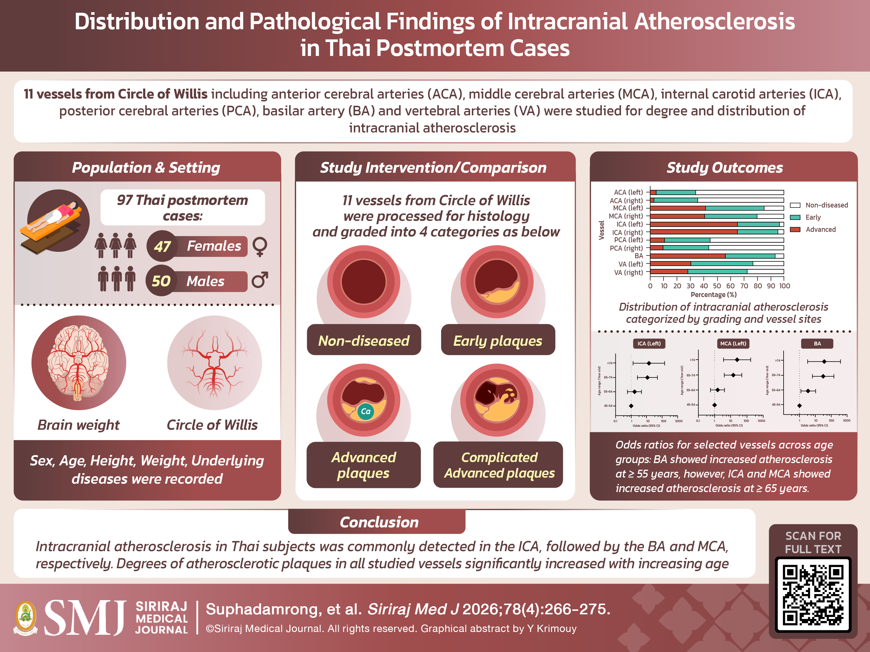
Objective: To determine the distribution and pathological findings of intracranial atherosclerosis in Thai postmortem cases.
Materials and Methods: The prospective cross-sectional study was conducted on Thai postmortem cases aged 45 years or older. Sex, age, weight, height, and brain weight were recorded for each case. Intracranial atherosclerosis was assessed in 11 vessels of the Circle of Willis (CoW), including the anterior cerebral arteries (ACA), middle cerebral arteries (MCA), internal carotid arteries (ICA), posterior cerebral arteries (PCA), basilar artery (BA) and vertebral arteries (VA), using histological examination. Grading of atherosclerotic plaques in each vessel was recorded. Descriptive statistics, bivariate correlation, and ordinal logistic regression were performed where appropriate.
Results: A total of 97 Thai subjects were recruited, consisting of 47 female and 50 male subjects, with a mean age of 61.41 years. Brain weights in male subjects were significantly higher than those in female subjects (p < 0.001). Brain weights in both female and male subjects were negatively correlated with increasing age (p < 0.001). Early and advanced atherosclerotic plaques were most frequently detected in the ICA, followed by the BA and MCA, respectively. Using ordinal logistic regression, it was found that degrees of intracranial atherosclerosis in all vessels of the CoW significantly increased with increasing age (p < 0.05).
Conclusion: Intracranial atherosclerosis in Thai subjects was commonly detected in the ICA, followed by the BA and MCA, respectively. The degrees of atherosclerotic plaques in all studied vessels significantly increased with increasing age.
References
Chantkran W, Chaisakul J, Rangsin R, Mungthin M, Sakboonyarat B. Prevalence of and factors associated with stroke in hypertensive patients in Thailand from 2014 to 2018: A nationwide cross-sectional study. Sci Rep. 2021;11(1):17614.
Suwanwela NC. Stroke epidemiology in Thailand. J Stroke. 2014;16(1):1-7.
Thanakiatpinyo T, Dajpratham P, Kovindha A, Kuptniratsaikul V. Quality of Life of Stroke Patients at One Year after Discharge from Inpatient Rehabilitation: A Multicenter Study. Siriraj Med J. 2021;73(4):216-23.
Ritz K, Denswil NP, Stam OC, van Lieshout JJ, Daemen MJ. Cause and mechanisms of intracranial atherosclerosis. Circulation. 2014;130(16):1407-14.
Banerjee C, Chimowitz MI. Stroke Caused by Atherosclerosis of the Major Intracranial Arteries. Circ Res. 2017;120(3):502-13.
Lee JH, Han SJ, Yun YH, Choi HC, Jung S, Cho SJ, et al. Posterior circulation ischemic stroke in Korean population. Eur J Neurol. 2006;13(7):742-8.
Keselman B, Gdovinová Z, Jatuzis D, Melo TPE, Vilionskis A, Cavallo R, et al. Safety and Outcomes of Intravenous Thrombolysis in Posterior Versus Anterior Circulation Stroke: Results From the Safe Implementation of Treatments in Stroke Registry and Meta-Analysis. Stroke. 2020;51(3):876-82.
Denswil NP, van der Wal AC, Ritz K, de Boer OJ, Aronica E, Troost D, et al. Atherosclerosis in the circle of Willis: Spatial differences in composition and in distribution of plaques. Atherosclerosis. 2016;251:78-84.
Llopis G, Quinones S, Konschake M, Simon De Blas C, Hernández LM, Abramovic A, et al. Atheromatosis of the brain-supplying arteries: Circle of Willis, basilar, vertebral and their branches. Ann Anat. 2022;243:151941.
Yang WJ, Wong KS, Chen XY. Intracranial Atherosclerosis: From Microscopy to High-Resolution Magnetic Resonance Imaging. J Stroke. 2017;19(3):249-60.
Du Bois D, Du Bois EF. A formula to estimate the approximate surface area if height and weight be known. Arch Internal Med. 1916;17:863–71.
Du Bois D, Du Bois EF. A formula to estimate the approximate surface area if height and weight be known. 1916. Nutrition. 1989;5(5):303-11; discussion 312-3.
Sheaff MT, Hopster DJ. Post-mortem technique handbook. 2nd ed. London:Springer;2005. p.283-85.
Virmani R, Kolodgie FD, Burke AP, Farb A, Schwartz SM. Lessons from sudden coronary death: a comprehensive morphological classification scheme for atherosclerotic lesions. Arterioscler Thromb Vasc Biol. 2000;20(5):1262-75.
Ferretti-Rebustini REL, Jacob-Filho W, Suemoto CK, Farfel JM, Leite REP, Grinberg LT, et al. Factors associated with morphometric brain changes in cognitively normal aging. Dement Neuropsychol. 2015;9(2):103-109.
Bell MD, Long T, Roden AC, Cooper FI, Sanchez H, Trower C, et al; Autopsy Committee of the College of American Pathologists. Updating Normal Organ Weights Using a Large Current Sample Database. Arch Pathol Lab Med. 2022;146(12):1486-1495.
Yang WJ, Wong KS, Chen XY. Intracranial Atherosclerosis: From Microscopy to High-Resolution Magnetic Resonance Imaging. J Stroke. 2017;19(3):249-260.
Jiang H, Ren K, Li T, Qian C, Gong S, Wang T, et al. Correlation of the characteristics of symptomatic intracranial atherosclerotic plaques with stroke types and risk of stroke recurrence: a cohort study. Ann Transl Med. 2022;10(12):658.
Published
How to Cite
License
Copyright (c) 2026 Siriraj Medical Journal

This work is licensed under a Creative Commons Attribution-NonCommercial-NoDerivatives 4.0 International License.
Authors who publish with this journal agree to the following conditions:
Copyright Transfer
In submitting a manuscript, the authors acknowledge that the work will become the copyrighted property of Siriraj Medical Journal upon publication.
License
Articles are licensed under a Creative Commons Attribution-NonCommercial-NoDerivatives 4.0 International License (CC BY-NC-ND 4.0). This license allows for the sharing of the work for non-commercial purposes with proper attribution to the authors and the journal. However, it does not permit modifications or the creation of derivative works.
Sharing and Access
Authors are encouraged to share their article on their personal or institutional websites and through other non-commercial platforms. Doing so can increase readership and citations.


