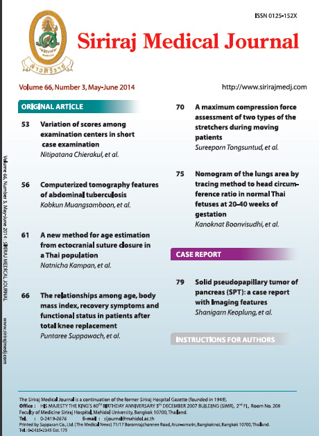
Solid Pseudopapillary Tumor of Pancreas (SPT): A Case Report with Imaging Features
Abstract
The solid pseudopapillary tumor of pancreas (SPT) is rare pancreatic tumor. There is a predilection for Asian and African-American women during the 2nd and 3rd decades of life. Clinical symptoms are usually slow-growing palpable mass, abdominal distension or incidentally discovered. Interestingly, diagnosis can be done with confidence by the classic imaging findings. In this report, we would like to present one case of young Thai female patient with the solid-cystic pancreatic mass demonstrating a well defined margin, internal hemorrhage and typical enhancing pattern.
Keywords: CT, pancreatic mass, pancreatic tumor, solid pseudopapillary tumor of pancreas
Siriraj Med J 2014;66:79-81
Downloads
Published
How to Cite
Issue
Section
License
Authors who publish with this journal agree to the following conditions:
Copyright Transfer
In submitting a manuscript, the authors acknowledge that the work will become the copyrighted property of Siriraj Medical Journal upon publication.
License
Articles are licensed under a Creative Commons Attribution-NonCommercial-NoDerivatives 4.0 International License (CC BY-NC-ND 4.0). This license allows for the sharing of the work for non-commercial purposes with proper attribution to the authors and the journal. However, it does not permit modifications or the creation of derivative works.
Sharing and Access
Authors are encouraged to share their article on their personal or institutional websites and through other non-commercial platforms. Doing so can increase readership and citations.


