
Isolated Unilateral Periorbital Chronic Cutaneous Lupus Erythematosus: A Case Report and Literature Review
Keywords:
Chronic Cutaneous Lupus Erythematosus, Periorbital Swelling, Chronic Cutaneous Lupus, Eyelid Dermatosis, Cutaneous Lupus ErythematosusAbstract
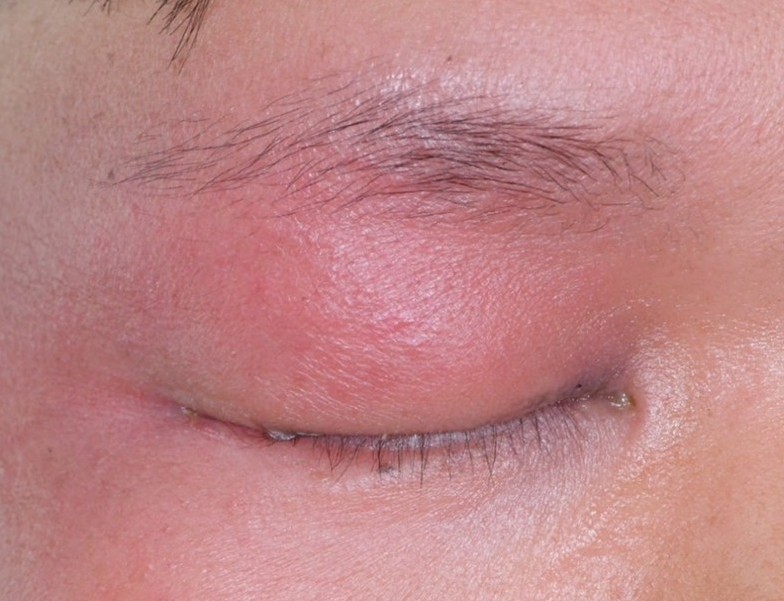
Chronic cutaneous lupus erythematosus (CCLE) represents the most prevalent subset of cutaneous lupus erythematosus and is well known for its predilection for photo-exposed areas, particularly the face and scalp, as consistently demonstrated in large clinical series and dermatologic epidemiologic studies. Periorbital involvement, particularly as an isolated manifestation, is exceedingly rare and diagnostically challenging. Here, we report a 29-year-old man presenting with a one-year history of recurrent isolated right periorbital swelling. Histopathological examination revealed interface dermatitis with basal vacuolization and lymphoplasmacytic infiltration, while Direct Immunofluorescent (DIF) showed granular deposition of C3 at the dermoepidermal junction. Despite negative antinuclear antibody (ANA) results, the patient responded favorably to hydroxychloroquine and topical corticosteroids. This case highlights the importance of maintaining a high clinical suspicion and utilizing DIF in diagnosing ANA-negative chronic cutaneous lupus erythematosus (CCLE) with atypical presentations.
References
Kuhn A, Landmann A. The classification and diagnosis of cutaneous lupus erythematosus. J Autoimmun 2014;48-49:14-9.
Bolognia JL, Schaffer JV, Cerroni L. Dermatology. 5th ed. Amsterdam: Elsevier 2024;p.670-88
Kang S, Amagai M, Bruckner AL, Enk AH, Margolis DJ, McMichael AJ, et al. Fitzpatrick’s Dermatology. 9th ed. McGraw Hill 2019;p.1037-1060
Elder DE, Elenitsas R, Johnson BL Jr, Murphy GF, Xu X. Lever’s Histopathology of the Skin. 12th ed. Philadelphia: Wolters Kluwer 2022;p.350-356
Makeeva V, Seminario-Vidal L, Beckum K, Sami N. Bilateral periorbital swelling as the initial presentation of cutaneous lupus erythematosus. JAAD Case Rep 2016;2:72-6.
Ghaninejad H, Kavusi S, Safar F, Asgari M, Naraghi ZS. Persistent periorbital edema as a sole manifestation of cutaneous lupus erythematosus: report of two cases. Dermatol Online J 2006;12:14.
Cyran S, Douglass MC, Silverstein JL. Chronic cutaneous lupus erythematosus presenting as periorbital edema and erythema. J Am Acad Dermatol 1992;26:334-8.
Bitar C, Menge TD, Chan MP. Cutaneous manifestations of lupus erythematosus: a practical clinicopathological review for pathologists. Histopathology 2022;81:669-88.
Schultz HY, Dutz JP, Furukawa F, Goodfield MJ, Kuhn A, Lee LA, et al. From pathogenesis, epidemiology, and genetics to definitions, diagnosis, and treatments of cutaneous lupus erythematosus and dermatomyositis: a report from the 3rd International Conference on Cutaneous Lupus Erythematosus (ICCLE) 2013. J Invest Dermatol 2015;135:7–12.
Wenzel J. Cutaneous lupus erythematosus: new insights into pathogenesis and therapeutic strategies. Nat Rev Rheumatol 2019;15:519-32.
Wallace DJ, Hahn BH, editors. Dubois’ lupus erythematosus and related syndromes. 10th ed. Philadelphia: Elsevier 2024;p. 738-49.
Lu Q, Long H, Chow S, Hidayat S, Danarti R, Listiawan Y, et al. Guideline for the diagnosis, treatment and long-term management of cutaneous lupus erythematosus. J Autoimmun 2021;123:102707.
Morand EF, Furie R, Tanaka Y, Bruce IN, Askanase AD, Richez C, et al. Trial of anifrolumab in active systemic lupus erythematosus. N Engl J Med 2020;382:211-21.
Gordon C, Merrill JT, Furie R, van Vollenhoven RF, Wallace DJ. Improvement in Cutaneous Lupus Erythematosus After Twenty Weeks of Belimumab Use: A Systematic Review and Meta-Analysis. Arthritis & Rheumatology 2022;74:1912-22.
Downloads
Published
How to Cite
Issue
Section
License
Copyright (c) 2026 Thai Journal of Dermatology

This work is licensed under a Creative Commons Attribution-NonCommercial-NoDerivatives 4.0 International License.
เนื้อหาและข้อมูลในบทความที่ลงตีพิมพ์ในวารสารโรคผิวหนัง ถือเป็นข้อคิดเห็นและความรับผิดชอบของผู้เขียนบทความโดยตรงซึ่งกองบรรณาธิการวารสาร ไม่จำเป็นต้องเห็นด้วย หรือร่วมรับผิดชอบใดๆ
บทความ ข้อมูล เนื้อหา รูปภาพ ฯลฯ ที่ได้รับการตีพิมพ์ในวารสารโรคผิวหนัง ถือเป็นลิขสิทธิ์ของวารสารฯ หากบุคคลหรือหน่วยงานใดต้องการนำทั้งหมดหรือส่วนหนึ่งส่วนใดไปเผยแพร่ต่อหรือเพื่อกระทำการใดๆ จะต้องได้รับอนุญาตเป็นลายลักอักษรจากบรรณาธิการวารสารโรคผิวหนังก่อนเท่านั้น

