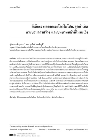
ดีเอ็นเอวงกลมนอกโครโมโซม: จุดกําเนิด กระบวนการสร้าง และบทบาทหน้าที่ในมะเร็ง
ดีเอ็นเอวงกลมนอกโครโมโซม
คำสำคัญ:
ดีเอ็นเอวงกลมนอกโครโมโซม, ยีนก่อมะเร็ง, ยีนดื้อยา, ตัวบ่งชี้ทางชีวภาพบทคัดย่อ
ดีเอ็นเอวงกลมนอกโครโมโซม (extrachromosomal circular DNA; eccDNA) เป็นดีเอ็นเอสายคู่ที่มีโครงสร้างเป็นวงกลม เกิดขึ้นจากความไม่เสถียรของจีโนม และดํารงอยู่แยกจากโครโมโซมอย่างอิสระ eccDNA มีขนาดที่หลากหลายและมีจุดกําเนิดที่กระจายอยู่ทั่วทั้งจีโนมสามารถตรวจพบได้ทั้งในเซลล์ปกติและเซลล์มะเร็ง อย่างไรก็ตามมีรายงานว่าปริมาณของ eccDNA ในเซลล์มะเร็งนั้นสูงกว่าซลล์ปกติอย่างมีนัยสําคัญ และที่น่าสนใจ คือ ระดับ eccDNA ที่เพิ่มขึ้นสามารถใช้เป็นตัวบ่งชี้พยาธิสภาพของโรคมะเร็งได้อย่างมีประสิทธิภาพ งานวิจัยได้รายงานการเพิ่มจํานวนของยีนก่อมะเร็งและยีนดื้อยาที่อยู่บนโครงสร้างของ eccDNA นั้น เป็นปัจจัยสําคัญในการขับเคลื่อนวิวัฒนาการและความหลากหลายทางพันธุกรรมของเนื้อเยื่อมะเร็ง รวมทั้งมีความสัมพันธ์กับภาวะดื้อยาและผลลัพธ์การพยากรณ์โรคที่ไม่ดี นอกจากนี้การศึกษาล่าสุดพบว่า eccDNA สามารถหลั่งออกนอกเซลล์เข้าสู่กระแสเลือด (cell-free eccDNA) และมีลักษณะทางพันธุกรรมที่เป็นเอกลักษณ์แตกต่างกันไปตามชนิดของเซลล์ รวมทั้งระดับการแสดงออกของยีนบน eccDNA ยังสัมพันธ์กับสถานะของโรคและอัตราการรอดชีพของผู้ป่วยอีกด้วย ดังนั้น eccDNA จึงมีแนวโน้มเป็นตัวบ่งชี้ทางชีวภาพที่มีศักยภาพสําหรับการติดตามผลการรักษาและการพยากรณ์โรคมะเร็ง บทความนี้จึงมีวัตถุประสงค์เพื่อนําเสนอความรู้ปัจจุบันเกี่ยวกับ eccDNA ที่ตรวจพบได้ทั้งในเนื้อเยื่อและกระแสเลือดของผู้ป่วยโรคมะเร็ง ตลอดจนแหล่งที่มา กลไกการเกิด และบทบาททางชีววิทยาที่สําคัญที่อาจนําไปสู่แนวทางการวินิจฉัยโรคที่แม่นยําและการรักษาโรคมะเร็งที่มีประสิทธิภาพยิ่งขึ้น
เอกสารอ้างอิง
Stanfield SW, Lengyel JA. Small circular DNA of Drosophila melanogaster: chromosomal homology and kinetic complexity. Proc Natl Acad Sci USA 1979; 76(12): 6142–6.
Møller HD, Parsons L, Jørgensen TS, Botstein D, Regenberg B. Extrachromosomal circular DNA is common in yeast. Proc Natl Acad Sci USA 2015; 112(24): E3114–22.
Møller HD, Mohiyuddin M, Prada-Luengo I, Sailani MR, Halling JF, Plomgaard P, et al. Circular DNA elements of chromosomal origin are common in healthy human somatic tissue. Nat Commun 2018; 9(1): 1069. (12 pages).
Kim H, Nguyen N-P, Turner K, Wu S, Gujar AD, Luebeck J, et al. Extrachromosomal DNA is associated with oncogene amplification and poor outcome across multiple cancers. Nat Genet 2020; 52(9): 891–7.
Wu S, Turner KM, Nguyen N, Raviram R, Erb M, Santini J, et al. Circular ecDNA promotes accessible chromatin and high oncogene expression. Nature 2019; 575(7784): 699–703.
Wu X, Li P, Yimiti M, Ye Z, Fang X, Chen P, et al. Identification and characterization of extrachromosomal circular DNA in plasma of lung adenocarcinoma patients. Int J Gen Med 2022; 15: 4781–91.
Kumar P, Dillon LW, Shibata Y, Jazaeri AA, Jones DR, Dutta A. Normal and cancerous tissues release extrachromosomal circular DNA (eccDNA) into the circulation. Mol Cancer Res 2017; 15(9): 1197–205.
Hoota Y, Bassel A. Molecular size and circularity of DNA in cells of mammals and higher plants. Proc Natl Acad Sci USA 1965; 53(2): 356–62.
Cox D, Yuncken C, Spriggs AI. Minute chromatin bodies in malignant tumours of childhood. Lancet 1965; 1(7402): 55–8.
Radloff R, Bauer W, Vinograd J. A dye-buoyant-density method for the detection and isolation of closed circular duplex DNA: the closed circular DNA in HeLa cells. Proc Natl Acad Sci USA 1967; 57(5): 1514–21.
Cohen S, Menut S, Méchali M. Regulated formation of extrachromosomal circular DNA molecules during development in Xenopus laevis. Mol Cell Biol 1999; 19(10): 6682–9.
Kunisada T, Yamagishi H, Ogita Z, Kirakawa T, Mitsui Y. Appearance of extrachromosomal circular DNAs during in vivo and in vitro ageing of mammalian cells. Mech Ageing Dev 1985; 29(1): 89–99.
Cohen S, Lavi S. Induction of circles of heterogeneous sizes in carcinogen-treated cells: two-dimensional gel analysis of circular DNA molecules. Mol Cell Biol 1996; 16(5): 2002–14.
Cohen S, Regev A, Lavi S. Small polydispersed circular DNA (spcDNA) in human cells: association with genomic instability. Oncogene 1997; 14(8): 977–85.
Schmidt H, Taubert H, Lange H, Kriese K, Schmitt WD, Hoff mann S, et al. Small polydispersed circular DNA contains strains of mobile genetic elements and occurs more frequently in permanent cell lines of malignant tumors than in normal lymphocytes. Oncol Rep 2009; 22(2): 393–400.
Kunisada T, Yamagishi H. Sequence organization of repetitive sequences enriched in small polydisperse circular DNAs from HeLa cells. J Mol Biol 1987; 198(4): 557–65.
Cohen S, Agmon N, Sobol O, Segal D. Extrachromosomal circles of satellite repeats and 5S ribosomal DNA in human cells. Mob DNA 2010; 1(1): 11. (11 pages).
Misra R, Matera AG, Schmid CW, Rush MG. Recombination mediates production of an extrachromosomal circular DNA containing a transposon-like human element, THE-1. Nucleic Acids Res 1989; 17(20): 8327–41.
Tomaska L, Nosek J, Kramara J, Griffith JD. Telomeric circles: universal players in telomere maintenance? Nat Struct Mol Biol 2009; 16(10): 1010–5.
Hull RM, Houseley J. The adaptive potential of circular DNA accumulation in ageing cells. Curr Genet 2020; 66(5): 889–94.
Dillon LW, Kumar P, Shibata Y, Wang YH, Willcox S, Griffith JD, et al. Production of extrachromosomal microDNAs is linked to mismatch repair pathways and transcriptional activity. Cell Rep 2015; 11(11): 1749–59.
Paulsen T, Shibata Y, Kumar P, Dillon L, Dutta A. Small extrachromosomal circular DNAs, microDNA, produce short regulatory RNAs that suppress gene expression independent of canonical promoters. Nucleic Acids Res 2019; 47(9): 4586–96.
Shoura MJ, Gabdank I, Hansen L, Merker J, Gotlib J, Levene SD, et al. Intricate and cell type-specific populations of endogenous circular DNA (eccDNA) in Caenorhabditis elegans and Homo sapiens. G3: Genes Genomes Genet 2017; 7(10): 3295–303.
Turner KM, Deshpande V, Beyter D, Koga T, Rusert J, Lee C, et al. Extrachromosomal oncogene amplification drives tumour evolution and genetic heterogeneity. Nature 2017; 543(7643): 122–5.
Chen YY, Dagg R, Zhang Y, Lee JHY, Lu R, Martin La Rotta N, et al. The C-Circle biomarker is secreted by alternative-lengthening-of-telomeres positive cancer cells inside exosomes and provides a blood-based diagnostic for ALT Activity. Cancers (Basel) 2021; 13(21). 5369. (24 pages).
Henson JD, Cao Y, Huschtscha LI, Chang AC, Au AYM, Pickett HA, et al. DNA C-circles are specific and quantifiable markers of alternative-lengthening-of-telomeres activity. Nat Biotechnol 2009; 27(12): 1181–5.
Luo X, Zhang L, Cui J, An Q, Li H, Zhang Z, et al. Small extrachromosomal circular DNAs as biomarkers for multi-cancer diagnosis and monitoring. Clin Transl Med 2023; 13(9): e1393. (16 pages).
Zakov S, Kinsella M, Bafna V. An algorithmic approach for breakage-fusion-bridge detection in tumor genomes. Proc Natl Acad Sci USA 2013; 110(14): 5546–51.
Mehanna P, Gagné V, Lajoie M, Spinella JF, St-Onge P, Sinnett D, et al. Characterization of the microDNA through the response to chemotherapeutics in lymphoblastoid cell lines. PloS one 2017; 12(9): e0184365. (14 pages).
Sunnerhagen P, Sjöberg RM, Karlsson AL, Lundh L, Bjursell G. Molecular cloning and characterization of small polydisperse circular DNA from mouse 3T6 cells. Nucleic Acids Res 1986; 14(20): 7823–38.
Huang C, Jia P, Chastain M, Shiva O, Chai W. The human CTC1/STN1/TEN1 complex regulates telomere maintenance in ALT cancer cells. Exp Cell Res 2017; 355(2): 95–104.
Shoshani O, Brunner SF, Yaeger R, Ly P, Nechemia-Arbely Y, Kim DH, et al. Chromo-thripsis drives the evolution of gene amplification in cancer. Nature 2021; 591(7848): 137–41.
Van Roy N, Vandesompele J, Menten B, Nilsson H, De Smet E, Rocchi M, et al. Translocationexcision-deletion-amplification mechanism leading to nonsyntenic coamplification of MYC and ATBF1. Genes Chromosom Cancer 2006; 45(2): 107–17.
Møller HD, Larsen CE, Parsons L, Hansen AJ, Regenberg B, Mourier T. Formation of extrachromosomal circular DNA from long terminal repeats of retrotransposons in Saccharomyces cerevisiae. G3: Genes Genomes Genet 2015; 6(2): 453–62.
Storlazzi CT, Lonoce A, Guastadisegni MC, Trombetta D, D'Addabbo P, Daniele G, et al. Gene amplification as double minutes or homogeneously staining regions in solid tumors: origin and structure. Genome Res 2010; 20(9): 1198–206.
Ling X, Han Y, Meng J, Zhong B, Chen J, Zhang H, et al. Small extrachromosomal circular DNA (eccDNA): major functions in evolution and cancer. Mol Cancer 2021; 20(1): 113. (15 pages).
Baker SP, Grant PA. The SAGA continues: expanding the cellular role of a transcriptional co-activator complex. Oncogene 2007; 26(37): 5329–40.
Cohen Z, Lavi S. Replication independent formation of extrachromosomal circular DNA in mammalian cell-free system. PLoS One 2009; 4(7): e6126. (8 pages).
Bailey C, Shoura MJ, Mischel PS, Swanton C. Extrachromosomal DNA-relieving heredity constraints, accelerating tumour evolution. Ann Oncol 2020; 31(7): 884–93.
deCarvalho AC, Kim H, Poisson LM, Winn ME, Mueller C, Cherba D, et al. Discordant inheritance of chromosomal and extrachromosomal DNA elements contributes to dynamic disease evolution in glioblastoma. Nat Genet 2018; 50(5): 708–17.
Lopez-Gines C, Gil-Benso R, Ferrer-Luna R, Benito R, Serna E, Gonzalez-Darder J, et al. New pattern of EGFR amplification in glioblastoma and the relationship of gene copy number with gene expression profile. Mod Pathol 2010; 23(6): 856–65.
Nguyen ND, Deshpande V, Luebeck J, Mischel PS, Bafna V. ViFi: accurate detection of viral integration and mRNA fusion reveals indiscriminate and unregulated transcription in proximal genomic regions in cervical cancer. Nucleic Acids Res 2018; 46(7): 3309–25.
Deshpande V, Luebeck J, Nguyen N-PD, Bakhtiari M, Turner KM, Schwab R, et al. Exploring the landscape of focal amplifications in cancer using AmpliconArchitect. Nat Commun 2019; 10(1): 392. (14 pages).
Jin K, Wang S, Zhang Y, Xia M, Mo Y, Li X, et al. Long non-coding RNA PVT1 interacts with MYC and its downstream molecules to synergistically promote tumorigenesis. Cell Mol Life Sci 2019; 76(21): 4275–89.
Chinen Y, Sakamoto N, Nagoshi H, Taki T, Maegawa S, Tatekawa S, et al. 8q24 amplified segments involve novel fusion genes between NSMCE2 and long noncoding RNAs in acute myelogenous leukemia. J Hematol Oncol 2014; 7: 68. (5 pages).
Koche RP, Rodriguez-Fos E, Helmsauer K, Burkert M, MacArthur IC, Maag J, et al. Extrachromosomal circular DNA drives oncogenic genome remodeling in neuroblastoma. Nat Genet 2020; 52(1): 29–34.
Del Rey J, Prat E, Ponsa I, Lloreta J, Gelabert A, Algaba F, et al. Centrosome clustering and cyclin D1 gene amplification in double minutes are common events in chromosomal unstable bladder tumors. BMC Cancer 2010; 10: 280. (11 pages).
Cooperman BS, Klinger HP. Double minute chromosomes in a case of acute myelogenous leukemia resistant to chemotherapy. Cytogenet Cell Genet 1981; 30(1): 25–30.
Amin AJ, Shaw M, Tadros J, Benn H, Maroules M. Double minute chromosome in acute myeloid leukemia. Blood 2006; 108(11): 4428.
Huennekens FM. The methotrexate story: a paradigm for development of cancer chemotherapeutic agents. Adv Enzyme Regul 1994; 34: 397–419.
Nathanson DA, Gini B, Mottahedeh J, Visnyei K, Koga T, Gomez G, et al. Targeted therapy resistance mediated by dynamic regulation of extrachromosomal mutant EGFR DNA. Science 2014; 343(6166): 72–6.
Xu K, Ding L, Chang TC, Shao Y, Chiang J, Mulder H, et al. Structure and evolution of double minutes in diagnosis and relapse brain tumors. Acta Neuropathol 2019; 137(1): 123–37.
Koduru P, Chen W, Haley B, Ho K, Oliver D, Wilson K. Cytogenomic characterization of double minute heterogeneity in therapy related acute myeloid leukemia. Cancer Genet 2019; 238: 69–75.
Xue Y, Martelotto L, Baslan T, Vides A, Solomon M, Mai TT, et al. An approach to suppress the evolution of resistance in BRAFV600E-mutant cancer. Nat Med 2017; 23(8): 929–37.
Su Z, Saha S, Paulsen T, Kumar P, Dutta A. ATAC-Seq-based Identification of extrachromosomal circular DNA in mammalian cells and its validation using inverse PCR and FISH. Bio Protoc 2021; 11(9): e4003. (21 pages).
Luebeck J, Ng AWT, Galipeau PC, Li X, Sanchez CA, Katz-Summercorn AC, et al. Extrachromosomal DNA in the cancerous transformation of Barrett’s oesophagus. Nature 2023; 616(7958): 798–805.
Mehta D, Cornet L, Hirsch-Hoffmann M, Zaidi SS-e-A, Vanderschuren H. Full-length sequencing of circular DNA viruses and extrachromosomal circular DNA using CIDER-Seq. Nat Protoc 2020; 15(5): 1673–89.
Chen JP, Diekmann C, Wu H, Chen C, Della Chiara G, Berrino E, et al. scCircle-seq unveils the diversity and complexity of extrachromosomal circular DNAs in single cells. Nat Commun 2024; 15(1): 1768. (14 pages).
Spain L, Coulton A, Lobon I, Rowan A, Schnidrig D, Shepherd STC, et al. Late-stage metastatic melanoma emerges through a diversity of evolutionary pathways. Cancer Discov 2023; 13(6): 1364–85.
Chang L, Deng E, Wang J, Zhou W, Ao J, Liu R, et al. Single-cell third-generation sequencingbased multi-omics uncovers gene expression changes governed by ecDNA and structural variants in cancer cells. Clin Transl Med 2023; 13(8): e1351. (23 pages).
Chen W, Weng Z, Xie Z, Xie Y, Zhang C, Chen Z, et al. Sequencing of methylase-accessible regions in integral circular extrachromosomal DNA reveals differences in chromatin structure. Epigenetics Chromatin 2021; 14: 40. (11 pages).
Lanciano S, Zhang P, Llauro C, Mirouze M. Identification of extrachromosomal circular forms of active transposable elements using mobilome-seq. Methods Mol Biol 2021; 2250: 87–93.
Chamorro González R, Conrad T, Stöber MC, Xu R, Giurgiu M, Rodriguez-Fos E, et al. Parallel sequencing of extrachromosomal circular DNAs and transcriptomes in single cancer cells. Nat Genet 2023; 55(5): 880–90.
Fan X, Yang C, Li W, Bai X, Zhou X, Xie H, et al. SMOOTH-seq: single-cell genome sequencing of human cells on a third-generation sequencing platform. Genome Biol 2021; 22(1): 195. (19 pages).
Raymond E, Faivre S, Weiss G, McGill J, Davidson K, Izbicka E, et al. Effects of hydroxyurea on extrachromosomal DNA in patients with advanced ovarian carcinomas. Clin Cancer Res 2001; 7(5): 1171–80.
Von Hoff DD, Waddelow T, Forseth B, Davidson K, Scott J, Wahl G. Hydroxyurea accelerates loss of extrachromosomally amplified genes from tumor cells. Cancer Res 1991; 51(23 Pt 1): 6273–9.
Sanchez AM, Barrett JT, Schoenlein PV. Fractionated ionizing radiation accelerates loss of amplified MDR1 genes harbored by extrachromosomal DNA in tumor cells. Cancer Res. 1998; 58(17): 3845–54.
Barreto SC, Uppalapati M, Ray A. Small circular DNAs in human pathology. Malays J Med Sci 2014; 21(3): 4–18.
ดาวน์โหลด
เผยแพร่แล้ว
รูปแบบการอ้างอิง
ฉบับ
ประเภทบทความ
สัญญาอนุญาต
ลิขสิทธิ์ (c) 2026 วารสารกรมวิทยาศาสตร์การแพทย์

อนุญาตภายใต้เงื่อนไข Creative Commons Attribution-NonCommercial-NoDerivatives 4.0 International License.