
การใช้ Monocyte Distribution Width (MDW) เพื่อบ่งชี้การติดเชื้อในกระแสเลือด
คำสำคัญ:
ติดเชื้อในกระแสเลือด, โมโนไซด์, ความไว, ความจำเพาะบทคัดย่อ
การติดเชื้อในกระแสเลือด (Sepsis) และภาวะช็อกจากการติดเชื้อ (Septic shock) เป็นสาเหตุการเสียชีวิตที่สำคัญในโรงพยาบาล การวินิจฉัย และให้การรักษาอย่างรวดเร็วสามารถลดอัตราตายของผู้ป่วย Biomarker ที่ใช้ในการวินิจฉัย Sepsis เช่น C-Reactive Protein (CRP) White blood cell count (WBC) และขนาดเซลล์โมโนไซต์ (Monocyte Distribution Width: MDW) ที่เพิ่มขึ้นใช้เป็นตัวชี้วัด Sepsis วัตถุประสงค์ของงานวิจัยครั้งนี้ เพื่อประเมินความสามารถ MDW ในการวินิจฉัย Sepsis โดยเก็บข้อมูลผู้ป่วยที่มีการตรวจ CBC MDW CRP และ Hemoculture ระหว่างเดือนตุลาคม 2564 ถึงกันยายน 2565 วิเคราะห์ด้วยสถิติ Diagnostic test และพื้นที่ใต้กราฟ (AUROC)
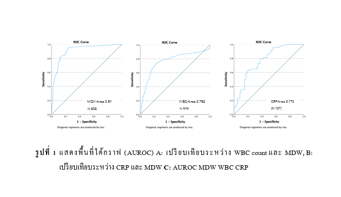
ข้อมูลผู้ป่วย 508 ราย พบ Hemoculture ผลบวก 295 ราย ผลลบ 213 ราย คิดเป็นร้อยละ 58.1 และ 41.9 กลุ่ม Hemoculture ผลบวกมีค่าเฉลี่ย CRP 138.4±135.9 mg/dl WBC 15,592.9±9,952.9 x103/ul MDW 30.7±10.9 AUROC ของ MDW CRP และ WBC เท่ากับ 0.910, 0.770 และ 0.762 ตามลำดับ Cut-off MDW 21.66 มีความไว ร้อยละ 87.46 ความจำเพาะ ร้อยละ 80.28 ค่าทำนายผลบวกร้อยละ 86.00 และค่าทำนายผลลบ ร้อยละ 82.21 พื้นที่ใต้กราฟระหว่าง MDW และ WBC พบว่า MDW มีความไวในการวินิจฉัย Sepsis มากกว่า WBC อย่างมีนัยสำคัญทางสถิติ (p < 0.001)
MDW เป็นพารามิเตอร์ที่มีประโยชน์ใช้วินิจฉัย Sepsis ได้จากการตรวจวิเคราะห์พื้นฐานทางห้องปฏิบัติการ การนำค่า MDW ประเมินร่วมกับข้อมูลการประเมินผู้ป่วยทางคลินิกสามารถเพิ่มความไว และความแม่นยำในการวินิจฉัย Sepsis
Downloads
เอกสารอ้างอิง
WHO. [Internet]. Available from: https://www.who.int/health-topics/sepsis#tab=tab_1. Sepsis. 2023.
กองยุทธศาสตร์และแผนงาน กระทรวงสาธารณสุข. รายละเอียดตัวชี้วัดกระทรวงสาธารณสุข ประจำปีงบประมาณ 2566. กรุงเทพมหานคร; 2566: 119–123.
Singer M, Deutschman CS, Seymour C, Shankar-Hari M, Annane D, Bauer M, et al. The third international consensus definitions for sepsis and septic shock (sepsis-3). JAMA - Journal of the American Medical Association. 2016; 315(8):801–10.
Thompson K, Venkatesh B, Finfer S. Sepsis and septic shock: current approaches to management. Intern Med J. 2019;49(2): 160–70.
Makic MBF, Bridges E. Managing Sepsis and Septic Shock: Current Guidelines and Definitions. American Journal of Nursing. 2018;118(2): 34–9.
Wile MJ, Homer LD, Gaehler S, Phillips S, Millan J. Manual differential cell counts help predict bacterial infection: A multivariate analysis. Am J Clin Pathol. 2001;115(5): 644–9.
Beckman Coulter. Early Sepsis Indicator (ESId) Application Addendum [Internet]. 2020. Available from: www.beckmancoulter.com/techdocs.
Crouser ED, Parrillo JE, Seymour C, Angus DC, Bicking K, Tejidor L, et al. Improved Early Detection of Sepsis in the ED With a Novel Monocyte Distribution Width Biomarker. Chest [Internet]. 2017;152(3):518–26. Available from: http://dx.doi.org/10.1016/j.chest.2017.05.039
Mukherjee R, Kanti Barman P, Kumar Thatoi P, Tripathy R, Kumar Das B, Ravindran B. Non-Classical monocytes display inflammatory features: Validation in Sepsis and Systemic Lupus Erythematous. Sci Rep. 2015; 5(July): 1–14.
Jo SJ, Kim SW, Choi JH, Choi SP, Lee J, Lim J. Monocyte distribution width (MDW) as a useful indicator for early screening of sepsis and discriminating false positive blood cultures. PLoS One. 2022; 17(12 December): 1–13.
Wu J, Li L, Luo J. Diagnostic and Prognostic Value of Monocyte Distribution Width in Sepsis. J Inflamm Res. 2022; 15(July): 4107–17.
Daniel WW. Biostatistics: A Foundation for Analysis in the Health Sciences. Vol. 44, Biometrics. 1988. 317 p.
Beckman Coulter. CRP Instructions For Use. Vol. May 2017. 2017. 1–8 p.
Malinovska A, Hinson JS, Badaki‐Makun O, Hernried B, Smith A, Debraine A, et al. Monocyte distribution width as part of a broad pragmatic sepsis screen in the emergency department. J Am Coll Emerg Physicians Open. 2022; 3(2): 1–11.
ดาวน์โหลด
เผยแพร่แล้ว
รูปแบบการอ้างอิง
ฉบับ
ประเภทบทความ
สัญญาอนุญาต
ลิขสิทธิ์ (c) 2023 สำนักงานสาธารณสุขจังหวัดกาฬสินธุ์

อนุญาตภายใต้เงื่อนไข Creative Commons Attribution-NonCommercial-NoDerivatives 4.0 International License.
เนื้อหาและข้อมูลในบทความที่ลงตีพิมพ์ในวารสารศูนย์ดัชนีการอ้างอิงวารสารไทย ถือเป็นข้อคิดเห็นและความรับผิดชอบของผู้เขียนบทความโดยตรงซึ่งกองบรรณาธิการวารสาร ไม่จำเป็นต้องเห็นด้วย หรือร่วมรับผิดชอบใด ๆบทความ ข้อมูล เนื้อหา รูปภาพ ฯลฯ ที่ได้รับการตีพิมพ์ในวารสารศูนย์ดัชนีการอ้างอิงวารสารไทย ถือเป็นลิขสิทธิ์ของวารสารศูนย์ดัชนีการอ้างอิงวารสารไทย หากบุคคลหรือหน่วยงานใดต้องการนำทั้งหมดหรือส่วนหนึ่งส่วนใดไปเผยแพร่ต่อหรือเพื่อกระทำการใด จะต้องได้รับอนุญาตเป็นลายลักณอักษรจากวารสารศูนย์ดัชนีการอ้างอิงวารสารไทยก่อนเท่านั้น


