
Amyloid PET Radiopharmaceuticals and Imaging for Clinical and Research Applications in Thailand
DOI:
https://doi.org/10.33192/smj.v75i9.263161Keywords:
Amyloid PET radiopharmaceuticals, Alzheimer’s disease, Amyloid beta plaque (Aβ), Molecular imaging, Dementia, amyloid depositionAbstract
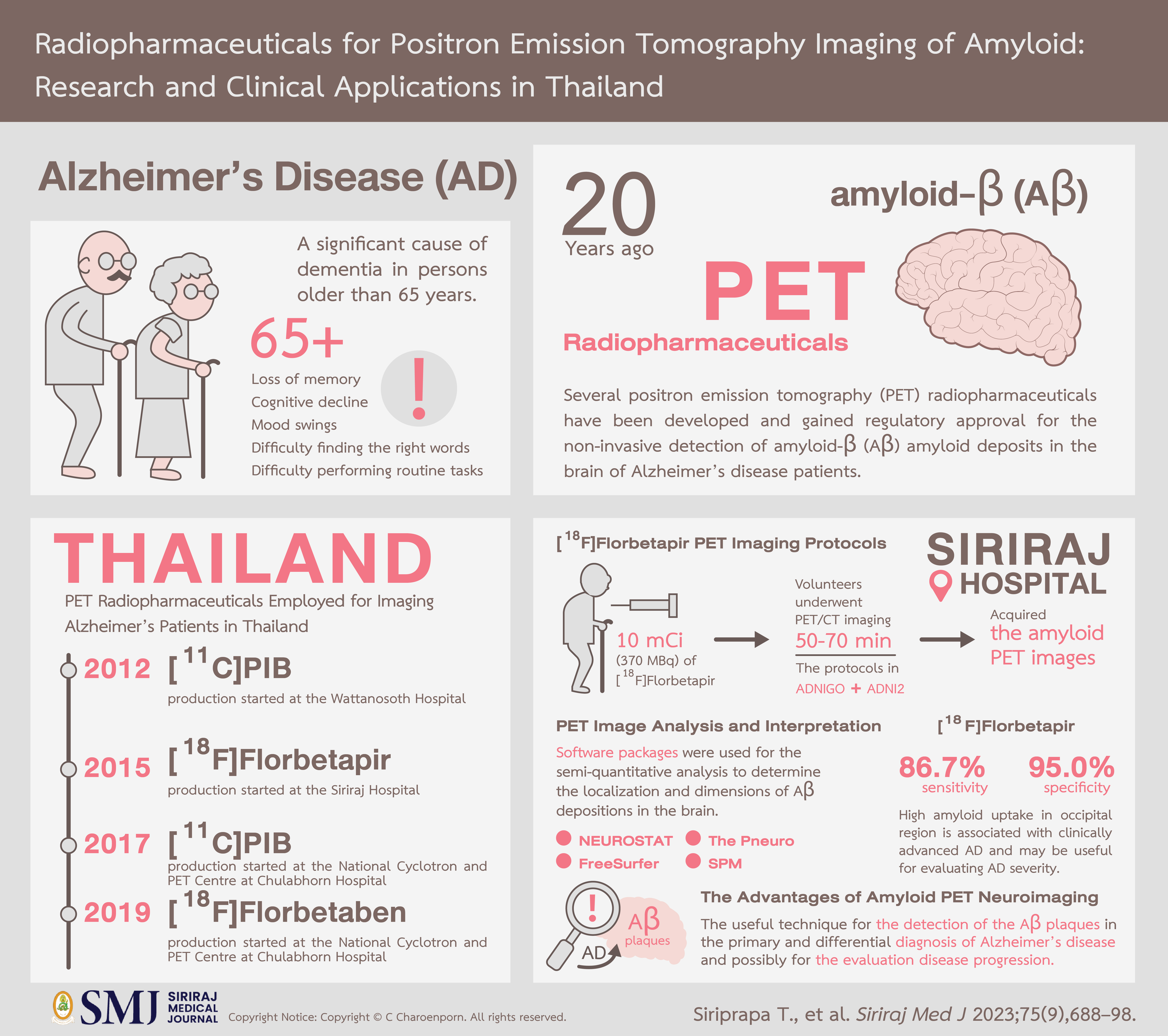
In the past two decades, the research community has focused on defining reliable molecular biomarkers for the early diagnosis of Alzheimer's disease (AD). Several PET radiopharmaceuticals have been developed and gained regulatory approval for the non-invasive detection of Aβ amyloid deposits in the brain. Nowadays, there are several PET imaging tracers available in Thailand for amyloid imaging including [11C]PiB, [18F]Florbetapir, and [18F]Florbetaben. This review provides a summary of commonly used amyloid PET radiopharmaceuticals, focusing on the available radiopharmaceuticals in Thailand and the experiences of using amyloid PET radiopharmaceuticals and imaging for clinical and research applications at Siriraj Hospital.
References
Maschio C, Ni R. Amyloid and tau positron emission tomography imaging in Alzheimer’s disease and other tauopathies. Front Aging Neurosci. 2022 [cited 2023 May 3];14:838034. Available from: https://pubmed.ncbi.nlm.nih.gov/35527737/
DeTure MA, Dickson DW. The neuropathological diagnosis of Alzheimer’s disease. Mol Neurodegener. 2019;14(1):32. Available from: http://dx.doi.org/10.1186/s13024-019-0333-5
About Alzheimer’s & dementia. Alzint.org. [cited 2023 May 3]. Available from: https://www.alzint.org/about/
Masters CL, Cappai R, Barnham KJ, Villemagne VL. Molecular mechanisms for Alzheimer’s disease: implications for neuroimaging and therapeutics. J Neurochem. 2006;97(6):1700–25. Available from: http://dx.doi.org/10.1111/j.1471-4159.2006.03989.x
Hu Z, Zeng L, Huang Z, Zhang J, Li T. The study of Golgi apparatus in Alzheimer’s disease. Neurochem Res. 2007 [cited 2023 May 3];32(8):1265–77. Available from: https://pubmed.ncbi.nlm.nih.gov/17401657/
Brookmeyer R, Johnson E, Ziegler-Graham K, Arrighi HM. Forecasting the global burden of Alzheimer’s disease. Alzheimers Dement. 2007 [cited 2023 May 3];3(3):186–91. Available from: https://pubmed.ncbi.nlm.nih.gov/19595937/
Leesri T. The study of prevalence and associated factors of dementia in the elderly. Siriraj Med J [Internet]. 2021 [cited 2023 Jun 1];73(4):224–35. Available from: https://he02.tci-thaijo.org/index.php/sirirajmedj/article/view/247824
Sukhatunga K, Phattarayuttawat S, Luchom M, Chantra J, Chaiyasit W, Bunnagulrote K. Depression and dementia in Thai elderly in urban and rural communities. Siriraj Med J [Internet]. 1999 [cited 2023 Jun 2];51(4):232–43. Available from: https://he02.tci-thaijo.org/index.php/sirirajmedj/article/view/246981
Senanarong V, Harnphadungkit K, Poungvarin N, Vannasaeng S, Chongwisal S, Chakorn T, et al. The Dementia and Disability Project in Thai Elderly: rational, design, methodology and early results. BMC Neurol. 2013 [cited 2023 May 3];13:3. Available from: https://pubmed.ncbi.nlm.nih.gov/23305293/
Tangwongchai S, Supasitthumrong T, Hemrunroj S, Tunvirachaisakul C, Chuchuen P, Houngngam N, et al. In Thai nationals, the ApoE4 allele affects multiple domains of neuropsychological, biobehavioral, and social functioning thereby contributing to Alzheimer’s disorder, while the ApoE3 allele protects against neuropsychiatric symptoms and psychosocial deficits. Mol Neurobiol. 2018 [cited 2023 May 3];55(8):6449–62. Available from: https://pubmed.ncbi.nlm.nih.gov/29307083/
Senanarong V, Siwasariyanon N, Washirutmangkur L, Poungvarin N, Ratanabunakit C, Aoonkaew N, et al. Alzheimer’s disease dementia as the diagnosis best supported by the cerebrospinal fluid biomarkers: difference in cut-off levels from thai experience. Int J Alzheimers Dis. 2012 [cited 2023 May 3];2012:212063. Available from: https://pubmed.ncbi.nlm.nih.gov/22844634/
Srisawat C, Junnu S, Peerapittayamongkol C, Futrakul A, Soi-ampornkul R, Senanarong V, et al. The platelet amyloid precursor protein ratio as a diagnostic marker for Alzheimer’s disease in Thai patients. J Clin Neurosci. 2013 [cited 2023 May 3];20(5):644–8. Available from: https://pubmed.ncbi.nlm.nih.gov/23453155/
Okamura N, Yanai K. Florbetapir (18F), a PET imaging agent that binds to amyloid plaques for the potential detection of Alzheimer’s disease. IDrugs [Internet]. 2010 [cited 2023 Jun 15];13(12):890–9. Available from: https://pubmed.ncbi.nlm.nih.gov/21154149/
Hyman BT. Amyloid-dependent and amyloid-independent stages of Alzheimer disease. Arch Neurol. 2011; 68:1062–1064.
Karran E, Mercken M, De Strooper B. The amyloid cascade hypothesis for Alzheimer’s disease: an appraisal for the development of therapeutics. Nat Rev Drug Discov. 2011;10:698–712.
Johnson KA, Gregas M, Becker JA, Kinnecom C, Salat DH, Moran EK, et al. Imaging of amyloid burden and distribution in cerebral amyloid angiopathy. Ann Neurol. 2007; 62:229–34.
Burack MA, Hartlein J, Flores HP, Taylor-Reinwald L, Perlmutter JS, Caims NJ. In vivo amyloid imaging in autopsy–confirmed Parkinson disease dementia. Neurology. 2010;74:77–84.
Rowe CC, Ng S, Ackermann U, Gong SJ, Pike K, Savage G, et al. Imaging beta-amyloid burden in aging and dementia. Neurology. 2007;68:1718–25.
Villemagne VL, McLean CA, Reardon K, Boyd A, Lewis V, Klug G, et al. 11C-PiB PET studies in typical sporadic Creutzfeldt–Jakob disease. J Neurol Neurosurg Psychiatry. 2009;80:998–1001.
Johansson A, Savitcheva I, Forsberg A, Engler H, Langstrom B, Nordberg A, et al. [(11)C]PIB imaging in patients with Parkinson’s disease. Preliminary results. Parkinsonism Relat Disord. 2008;14:345–7.
Landau SM, Thomas BA, Thurfjell L, Schmidt M, Margolin R, Mintun M, et al. Amyloid PET imaging in Alzheimer’s disease: a comparison of three radiotracers. Eur J Nucl Med Mol Imaging. 2014;41(7):1398-407.
Wong DF, Rosenberg PB, Zhou Y, Kumar A, Raymont V, Ravert HT, et al. In vivo imaging of amyloid deposition in Alzheimer’s disease using the radioligand 18F-AV-45 (florbetapir[corrected] F 18). J Nucl Med. 2010;51(6):913–20.
Clark CM, Pontecorvo MJ, Beach TG, Bedell BJ, Coleman RE, Doraiswamy PM, et al. Cerebral PET with florbetapir compared with neuropathology at autopsy for detection of neuritic amyloid-beta plaques: a prospective cohort study. Lancet Neurol. 2012;11(8):669–78.
Sabri O, Sabbagh MN, Seibyl J, Barthel H, Akatsu H, Ouchi Y, et al. Florbetaben PET imaging to detect amyloid beta plaques in Alzheimer’s disease: phase 3 study. Alzheimers Demen. 2015;11(8):964–74.
Villemagne, VL, Mulligan RS, Pejoska S, Ong K, Jones G, O’Keefe G, et al. Comparison of (11)C-PiB and (18)F-florbetaben for A imaging in ageing and Alzheimer’s disease. Eur J Nucl Med Mol Imaging. 2012; 39(6):983–9.
Biancalana M, Koide S. Molecular mechanism of Thioflavin-T binding to amyloid fibrils. Biochim Biophys Acta. 2010 [cited 2023 May 3];1804(7):1405–12. Available from: https://pubmed.ncbi.nlm.nih.gov/20399286/
Yakupova EI, Bobyleva LG, Vikhlyantsev IM, Bobylev AG. Congo Red and amyloids: history and relationship. Biosci Rep. 2019 [cited 2023 May 3];39(1):BSR20181415. Available from: https://pubmed.ncbi.nlm.nih.gov/30567726/
Klunk WE, Debnath ML, Pettegrew JW. Chrysamine-G binding to Alzheimer and control brain: an autopsy study of a new amyloid probe. Neurobiol Aging. 1995 [cited 2023 May 3];16(4):541–8. Available from: https://pubmed.ncbi.nlm.nih.gov/8544903/
Benveniste H, Einstein G, Kim KR, Hulette C, Johnson GA. Detection of neuritic plaques in Alzheimer’s disease by magnetic resonance microscopy. Proc Natl Acad Sci USA. 1999;96(24):14079–84.
Agdeppa ED, Kepe V, Liu J, Flores-Torres S, Satyamurthy N, Petric A, et al. Binding characteristics of radiofluorinated 6-dialkylamino-2-naphthylethylidene derivatives as positron emission tomography imaging probes for ß-amyloid plaques in Alzheimer’s disease. J Neurosci. 2001;21(24):RC189.
Mathis CA, Bacskai BJ, Kajdasz ST, McLellan ME, Frosch MP, Hyman BT, et al. A lipophilic thioflavin-T derivative for positron emission tomography (PET) imaging of amyloid in the brain. Bioorg Med Chem Lett. 2002;12(3):295-8.
Mallik A, Drzezga A, Minoshima S. Clinical amyloid imaging. Semin Nucl Med. 2017;47(1):31–43.
Rowe CC, Ackerman U, Browne W, Mulligan R, Pike KL, O’Keefe G, et al. Imaging of amyloid beta in Alzheimer’s disease with(18)F-BAY94-9172, a novel PET tracer: proof of mechanism. Lancet Neurol. 2008;7(2):129–35.
Serdons K, Terwinghe C, Vermaelen P, Van Laere K, Kung H, Mortelmans L, et al. Synthesis and evaluation of (18)F-labeled 2-phenylbenzothiazoles as positron emission tomography imaging agents for amyloid plaques in Alzheimer’s disease. J Med Chem. 2009;52(5):1428-37.
Wong DF, Rosenberg PB, Zhou Y, Kumar A, Raymont V, Ravert HT, et al. In vivo imaging of amyloid deposition in Alzheimer’s disease using the radioligand 18F-AV-45 (florbetapir[corrected] F 18). J Nucl Med. 2010;51(6):913–20.
Rowe CC, Jones G, Doré V, Pejoska S, Margison L, Mulligan RS, et al. Standardized expression of 18F-NAV4694 and 11C-PiB β-amyloid PET results with the centiloid scale. J Nucl Med [Internet]. 2016 [cited 2023 Jun 20];57(8):1233-7. Available from: https://pubmed.ncbi.nlm.nih.gov/26912446/
Cselényi Z, Jönhagen ME, Forsberg A, Halldin C, Julin P, Schou M, et al. Clinical validation of 18F-AZD4694, an Aβ-specific PET radioligand. J Nucl Med. 2012;53(3):415–24.
Thompson PW, Ye L, Morgenstern JL, Sue L, Beach TG, Judd DJ, et al. Interaction of the amyloid imaging tracer FDDNP with hallmark Alzheimer’s disease pathologies. J Neurochem. 2009;109:623–30.
Cohen AD, Rabinovici GD, Mathis CA, Jagust WJ, Klunk WE, Ikonomovic MD. Using Pittsburgh Compound B for in vivo PET imaging of fibrillar amyloid-beta. Adv Pharmacol. 2012;64:27–81.
Zhang W, Oya S, Kung MP, Hou C, Maier DL, Kung HF. F-18 stilbenes as PET imaging agents for detecting beta-amyloid plaques in the brain. J Med Chem. 2005;48(19):5980–8.
Cselenyi Z, Jonhagen ME, Forsberg A, Halldin C, Julin P, Schou M, et al. Clinical validation of 18F-AZD4694, an amyloid-beta-specific PET radioligand. J Nucl Med. 2012;53(3):415–24.
Rowe CC, Pejoska S, Mulligan RS, Jones G, Chan JG, Svensson S, et al. Head-to-head comparison of 11C-PiBand 18F-AZD4694 (NAV4694) for beta-amyloid imaging in aging and dementia. J Nucl Med. 2013;54(6):880–6.
Grimmer T, Shi K, Diehl-Schmid J, Natale B, Drzezga A, Förster S, et al. 18F-FIBT may expand PET for β-amyloid imaging in neurodegenerative diseases. Mol Psychiatry. 2020 [cited 2023 May 16];25(10):2608–19. Available from: https://pubmed.ncbi.nlm.nih.gov/30120417/
Yousefi BH, von Reutern B, Scherübl D, Manook A, Schwaiger M, Grimmer T, et al. FIBT versus florbetaben and PiB: a preclinical comparison study with amyloid-PET in transgenic mice. EJNMMI Res [Internet]. 2015 [cited 2023 Jun 20];5(1):20. Available from: https://pubmed.ncbi.nlm.nih.gov/25918674/
Ruangma A, Panpitpat S, Saonam T, Kijprayoon S, Ngokpol S, Tanasirimanon M, et al. Challenges in Production of Alzheimer’s Tracer C-11 PiB. Bangk Med J. 2015 [cited 2023 May 3];09(01):70–5. Available from: https://he02.tci-thaijo.org/index.php/bkkmedj/article/view/221103
Chotipanich C, Promteangtrong C, Kunawudhi A. Development of 18F-FLT, 11C-PiB, 18F-THK 5351, and 68Ga-PSMA at the National Cyclotron and PET Centre, Chulabhorn Royal Academy. J Med Assoc Thailand. 2018 [cited 2023 May 3];101(6):199. Available from: http://www.jmatonline.com/index.php/jmat/article/view/9499
Thientunyakit T, Sethanandha C, Muangpaisan W, Chawalparit O, Arunrungvichian K, Siriprapa T, et al. Relationships between amyloid levels, glucose metabolism, morphologic changes in the brain, and clinical status of patients with Alzheimer’s disease. Ann Nucl Med. 2020;34(5):337–48.
Zhang W, Oya S, Kung M-P, Hou C, Maier DL, Kung HF. F-18 Polyethyleneglycol stilbenes as PET imaging agents targeting Abeta aggregates in the brain. Nucl Med Biol. 2005;32(8):799–809.
Liu Y, Zhu L, Plössl K, Choi SR, Qiao H, Sun X, et al. Optimization of automated radiosynthesis of [18F]AV-45: a new PET imaging agent for Alzheimer’s disease. Nucl Med Biol. 2010;37(8):917–25.
Cohen AD, Klunk WE. Early detection of Alzheimer’s disease using PiB and FDG PET. Neurobiol Dis [Internet]. 2014 [cited 2023 Jun 15];72 Pt A:117–22. Available from: http://dx.doi.org/10.1016/j.nbd.2014.05.001
Alzheimer’s Disease Neuroimaging Initiative website. ADNI-GO PET technical procedures manual: FDG & AV-45. https:/adni.loni.usc.edu/wpcontent/uploads/2010/05/adni2_pet_tech_manual_0142011.pdf.
Alzheimer’s Disease Neuroimaging Initiative website. ADNI 2 PET technical procedures manual: Florbetapir. Available at: https:/adni.loni.usc.edu/wp-content/uploads/2010/05/ADNI2 PET Tech Manual 0142011.pdf
Thientunyakit T, Thongpraparn T, Sethanandha C, Yamada T, Kimura Y, Muangpaisan W, et al. Relationship between F-18 florbetapir uptake in the occipital lobe and neurocognitive performance in Alzheimer’s disease. Jpn J Radiol. 2021;39(10):984–93.
Thientunyakit T, Sethanandha C, Muangpaisan W, Minoshima S. 3D-SSP analysis for amyloid brain PET imaging using 18F-florbetapir in patients with Alzheimer’s dementia and mild cognitive impairment. Med J Malaysia. 2021;76(4):493–501.
Wongsawaeng D, Chawalparit O, Piyapittayanan S, Thientunyakit T, Muangpaisan W, Thana-udom K, et al. Magnetic resonance hippocampal subfield volumetric analysis for differentiating among healthy older adults and older adults with mild cognitive impairment or major depressive disorder. Siriraj Med J [Internet]. 2021 [cited 2023 Jun 1];73(12):786–92. Available from: https://he02.tci-thaijo.org/index.php/sirirajmedj/article/view/254580
Minoshima S, Drzezga AE, Barthel H, Bohnen N, Djekidel M, Lewis DH, et al. SNMMI procedure standard/EANM practice guideline for amyloid PET imaging of the brain 1.0. J Nucl Med. 2016;57(8):1316–22.
PMOD Neuro Tool (PNEURO). Pmod.com. [cited 2023 May 16]. Available from: http://www.pmod.com/files/download/v35/doc/PDF/PNEURO.pdf
Desikan RS, Ségonne F, Fischl B, Quinn BT, Dickerson BC, Blacker D, et al. An automated labeling system for subdividing the human cerebral cortex on MRI scans into gyral-based regions of interest. Neuroimage. 2006;31(3):968–80.
Thientunyakit T, Sethanandha C, Muangpaisan W, Chawalparit O, Arunrungvichian K, Siriprapa T, et al. Implementation of [18F]-labeled amyloid brain PET imaging biomarker in the diagnosis of Alzheimer’s disease: first-hand experience in Thailand: First-hand experience in Thailand. Nucl Med Commun. 2018;39(2):186–92.
Jack CR Jr, Bennett DA, Blennow K, Carrillo MC, Dunn B, Haeberlein SB, et al. NIA-AA Research Framework: Toward a biological definition of Alzheimer’s disease. Alzheimer’s Dement. 2018 [cited 2023 May 16];14(4):535–62. Available from: https://psycnet.apa.org/fulltext/2018-15859-012.pdf
Thientunyakit T, Shiratori S, Ishii K, Gelovani JG. Molecular PET imaging in Alzheimer’s disease. J Med Biol Eng. 2022;42(3):301–17.
Johnson KA, Sperling RA, Gidicsin CM, Carmasin JS, Maye JE, Coleman RE, et al. Florbetapir (F18‐AV‐45) PET to assess amyloid burden in Alzheimer’s disease dementia, mild cognitive impairment, and normal aging. Alzheimer’s Dement. 2013;9(5S):S72–83.
Minoshima S, Drzezga AE, Barthel H, Bohnen N, Djekidel M, Lewis DH, et al. SNMMI procedure standard/EANM practice guideline for amyloid PET imaging of the brain 1.0. J Nucl Med. 2016;57(8):1316–22.
Chanisa C, Monchaya N, Anchisa K, Chetsadaporn P, Attapon J. Analysis of amyloid and tau deposition in Alzheimer’s disease using 11C-Pittsburgh compound B and 18F-THK 5351 positron emission tomography imaging. World J Nucl Med [Internet]. 2021;20(1):61–72. Available from: http://dx.doi.org/10.4103/wjnm.
Promteangtrong C, Poenateetai C, Jantarato A, Boonsingma N, Kunawudhi A, Chotipanich C. Impact of molecular imaging on the diagnosis of dementia subtypes. J Med Assoc Thai [Internet]. 2021 [cited 2023 Jul 18];104(12):1873–80.
Published
How to Cite
License
Copyright (c) 2023 Siriraj Medical Journal

This work is licensed under a Creative Commons Attribution-NonCommercial-NoDerivatives 4.0 International License.
Authors who publish with this journal agree to the following conditions:
Copyright Transfer
In submitting a manuscript, the authors acknowledge that the work will become the copyrighted property of Siriraj Medical Journal upon publication.
License
Articles are licensed under a Creative Commons Attribution-NonCommercial-NoDerivatives 4.0 International License (CC BY-NC-ND 4.0). This license allows for the sharing of the work for non-commercial purposes with proper attribution to the authors and the journal. However, it does not permit modifications or the creation of derivative works.
Sharing and Access
Authors are encouraged to share their article on their personal or institutional websites and through other non-commercial platforms. Doing so can increase readership and citations.


