
The Refractive Legacy of Prematurity: A Systematic Review and Meta-Analysis of Refractive Errors and Ocular Biometry in Infants With and Without Retinopathy of Prematurity
DOI:
https://doi.org/10.33192/smj.v78i7.282004Keywords:
Retinopathy of prematurity, Preterm, Myopia, Hyperopia, Astigmatism, Meta-analysis, Ocular biometryAbstract
Objective: To compare the prevalence of refractive errors in children born prematurely with and without retinopathy of prematurity (ROP) and to identify potential factors contributing to heterogeneity.
Materials and Methods: Relevant literature was systematically searched in PubMed, EMBASE, and CINAHL for studies available through January 2025. Studies were eligible if they used cross-sectional, cohort, or case-control designs and reported refractive outcomes among preterm children stratified by ROP status. Effect estimates were synthesized using random-effects models and expressed as pooled odds ratios and mean differences. Between-study variability was evaluated using Cochran’s Q statistic and the I² metric. Additional analyses included subgroup comparisons, meta-regression, and assessment of potential publication bias.
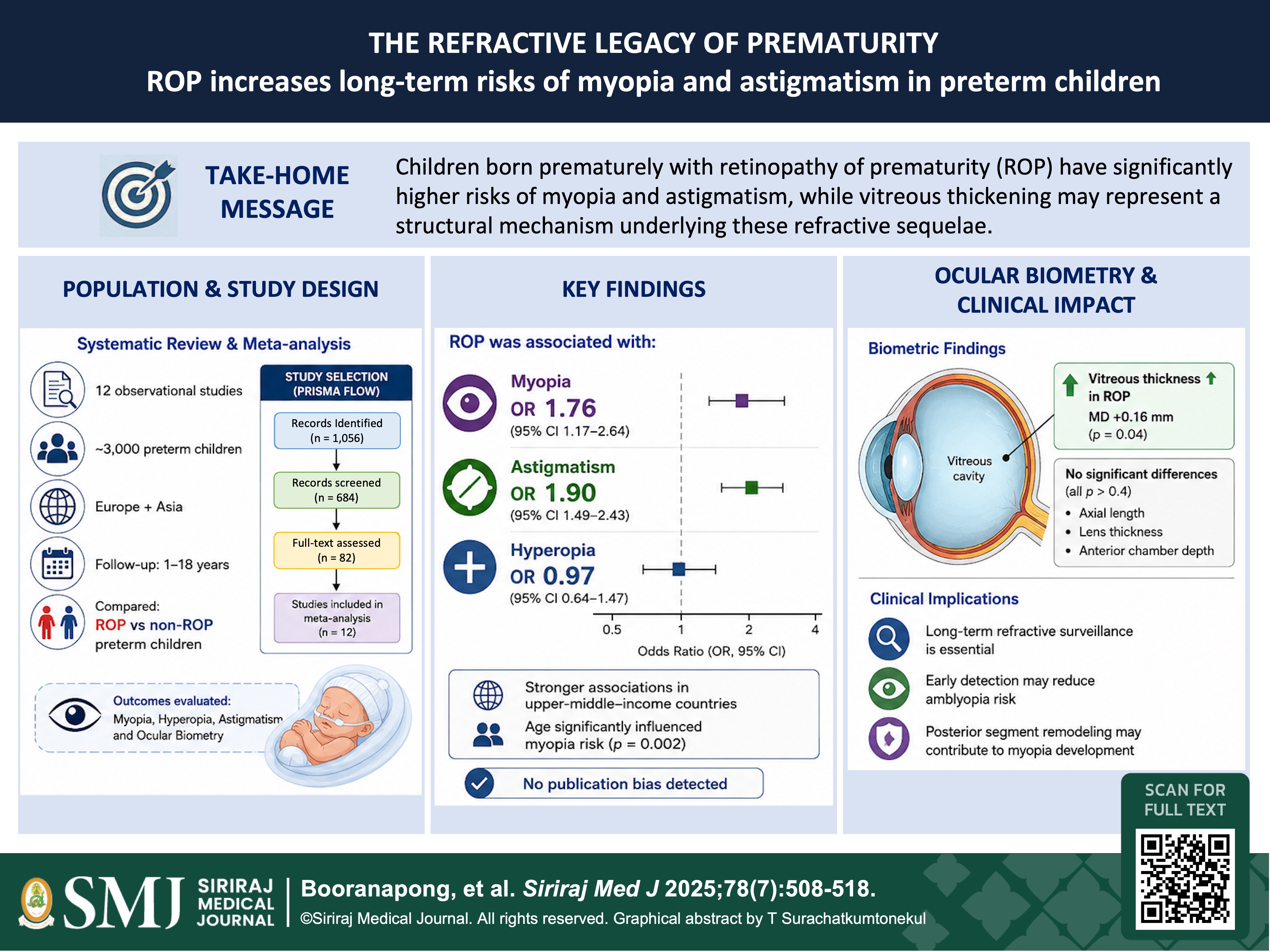
Results: Twelve studies were included. ROP was significantly associated with myopia (OR, 1.76; 95% CI, 1.17–2.64; p = 0.01) and astigmatism (OR, 1.90; 95% CI, 1.49–2.43; p < 0.001), but not hyperopia (OR, 0.97; 95% CI, 0.64–1.47; p = 0.89). Strong associations were observed in upper-middle–income countries. Age significantly modified the ROP–myopia association (p = 0.002). Biometric analysis showed greater vitreous thickness in ROP children (mean difference, 0.16 mm; p = 0.04), while axial length, lens thickness, and anterior chamber depth were not significantly
different (all p > 0.4). No publication bias was detected.
Conclusions: ROP is associated with increased risks of myopia and astigmatism, with effects influenced by age and socioeconomic context. Vitreous thickness was the only biometric parameter significantly affected, suggesting a potential mechanism underlying refractive sequelae, although this requires further validation.
References
Blencowe H, Cousens S, Chou D, Oestergaard M, Say L, Moller AB, et al. Born too soon: the global epidemiology of 15 million preterm births. Reproductive Health. 2013;10(Suppl 1):S2.
Stoll BJ, Hansen NI, Bell EF, Walsh MC, Carlo WA, Shankaran S, et al. Trends in Care Practices, Morbidity, and Mortality of Extremely Preterm Neonates, 1993-2012. JAMA. 2015;314(10):1039-51.
O’Connor AR, Stephenson TJ, Johnson A, Tobin MJ, Ratib S, Fielder AR. Long-term ophthalmic outcome of low birth weight children with and without retinopathy of prematurity. Pediatrics. 2006;118(6):2329–39.
Sirivunnabood T, Wanitpongpan P, Yapan P. Incidence and risk factors of neonatal sepsis in preterm premature rupture of membranes before 34 weeks of gestation. Siriraj Med J. 2022;74(3):169-77.
Quinn GE. Retinopathy of prematurity blindness worldwide: phenotypes in the third epidemic. Eye Brain. 2016;8:31-36.
Gilbert C, Foster A. Childhood blindness in the context of VISION 2020—the right to sight. Bull World Health Organ. 2001;79(3):227–32.
Phruksarudee K, Sungprem K, Montriwet M. Association of oxygen therapy concentration and duration with retinopathy of prematurity incidence at Naresuan University Hospital. Siriraj Med J. 2024;76(3):160-6.
Holmström GE, Larsson EK. Development of spherical equivalent refraction in prematurely born children during the first 10 years of life: a population-based study. Arch Ophthalmol. 2005;123(10):1404–11.
Larsson EK, Rydberg AC, Holmström GE. A population-based study of the refractive outcome in 10-year-old preterm and full-term children. Arch Ophthalmol. 2003;121(10):1430–6.
Cook A, White S, Batterbury M, Clark D. Ocular growth and refractive error development in premature infants with or without retinopathy of prematurity. Invest Ophthalmol Vis Sci. 2008;49(12):5199-207.
Cauduro R, de Moraes NSB, Goulart AL, Allemann N. Ocular biometry in preterm newborns in the neonatal intensive care unit environment of a referral hospital. Ophthalmol Sci. 2025;5(6):100829.
Webber AL, Wood J. Amblyopia: prevalence, natural history, functional effects and treatment. Clin Exp Optom. 2005;88(6):365–75.
Wang Y, Pi LH, Zhao RL, Zhu XH, Ke N. Refractive status and optical components of premature babies with or without retinopathy of prematurity at 7 years old. Transl Pediatr. 2020;9(2):108–16.
Chen TC, Tsai TH, Shih YF, Yeh PT, Yang CH, Hu FC, et al. Long-term evaluation of refractive status and optical components in eyes of children born prematurely. Invest Ophthalmol Vis Sci. 2010;51(12):6140–8.
Al Oum M, Donati S, Cerri L, Agosti M, Azzolini C. Ocular alignment and refraction in preterm children at 1 and 6 years old. Clin Ophthalmol. 2014;8:1263–8.
Zhu X, Zhao R, Wang Y, Ouyang L, Yang J, Li Y, et al. Refractive state and optical compositions of preterm children with and without retinopathy of prematurity in the first 6 years of life. Medicine (Baltimore). 2017;96(45):e8565.
Fieß A, Kölb-Keerl R, Schuster AK, Knuf M, Kirchhof B, Muether PS, et al. Prevalence and associated factors of strabismus in former preterm and full-term infants between 4 and 10 years of age. BMC Ophthalmol. 2017;17:228.
Rasoulinejad SA, Pourdad P, Pourabdollah A, Arzani A, Geraili Z, Roshan HY. Ophthalmologic outcome of premature infants with or without retinopathy of prematurity at 5–6 years of age. J Family Med Prim Care. 2020;9(9):4582–6.
Xie X, Wang Y, Zhao R, Yang J, Zhu X, Ouyang L, et al. Refractive status and optical components in premature infants with and without retinopathy of prematurity: a 4- to 5-year cohort study. Front Pediatr. 2022;10:922303.
Wang Y, Pi L, Zhao R, Zhu X, Ke N. Refractive status and optical components of premature babies with or without retinopathy of prematurity: a long-term study. Front Pediatr. 2022;10:922303.
Pétursdóttir D, Holmström G, Larsson E. Refraction and its development in young adults born prematurely and screened for retinopathy of prematurity. Acta Ophthalmol. 2022;100(2):189–95.
Genc CD, Yucel OE. Effects of prematurity and retinopathy of prematurity on refractive errors and biometric optic components in school children: results of a tertiary center from Turkey. Int Ophthalmol. 2023;43(11):4821–30.
Saluja G, Kaur K. Childhood Myopia and Ocular Development [Internet]. In: StatPearls [Internet]. Treasure Island (FL): StatPearls Publishing; 2025 Jan– [updated 2023 May 4; cited YEAR MONTH DAY]. Available from: https://www.ncbi.nlm.nih.gov/books/NBK587350/
Wallman J, Winawer J. Homeostasis of eye growth and the question of myopia. Neuron. 2004;43(4):447–68.
Flitcroft DI. The complex interactions of retinal, optical and environmental factors in myopia aetiology. Prog Retin Eye Res. 2012;31(6):622–60.
Schaeffel F, Wildsoet C. Can the retina alone detect the sign of defocus? Ophthalmic Physiol Opt. 2013;33(3):362–7.
Published
How to Cite
License
Copyright (c) 2026 Siriraj Medical Journal

This work is licensed under a Creative Commons Attribution-NonCommercial-NoDerivatives 4.0 International License.
Authors who publish with this journal agree to the following conditions:
Copyright Transfer
In submitting a manuscript, the authors acknowledge that the work will become the copyrighted property of Siriraj Medical Journal upon publication.
License
Articles are licensed under a Creative Commons Attribution-NonCommercial-NoDerivatives 4.0 International License (CC BY-NC-ND 4.0). This license allows for the sharing of the work for non-commercial purposes with proper attribution to the authors and the journal. However, it does not permit modifications or the creation of derivative works.
Sharing and Access
Authors are encouraged to share their article on their personal or institutional websites and through other non-commercial platforms. Doing so can increase readership and citations.