
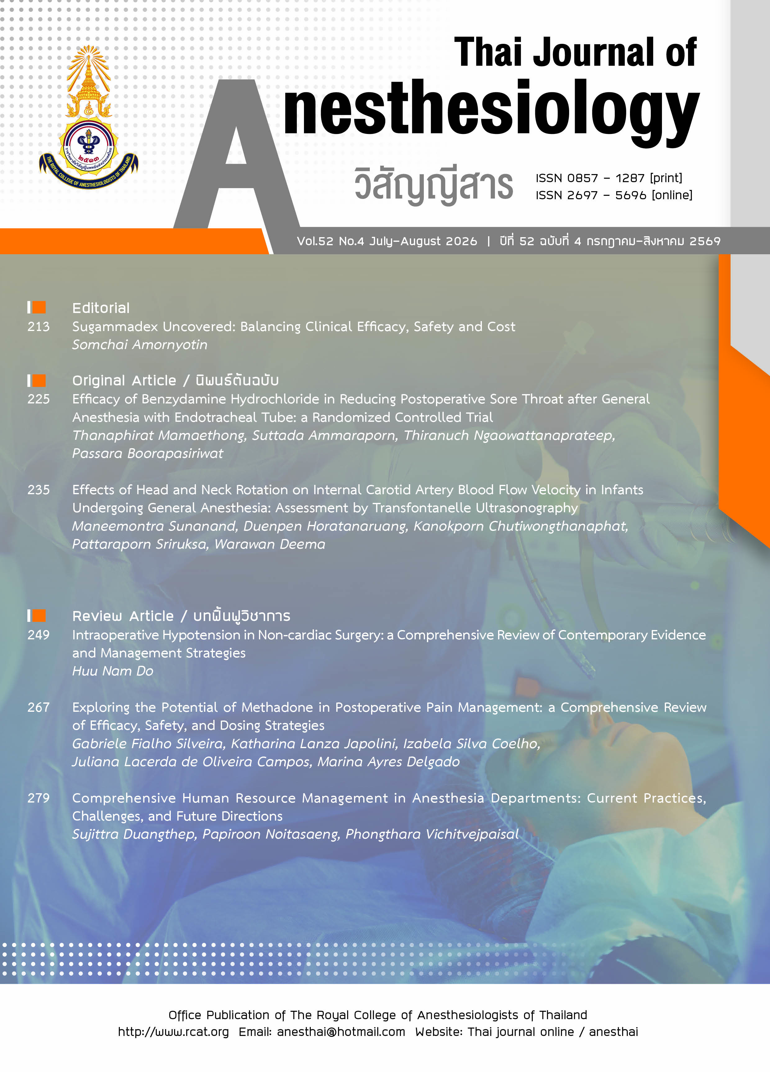
Effects of Head and Neck Rotation on Internal Carotid Artery Blood Flow Velocity in Infants Undergoing General Anesthesia: assessment by Transfontanelle Ultrasonography
Keywords:
cerebral blood flow velocity, head and neck rotation, infant, pediatric anesthesia, transfontanelle doppler ultrasonographyAbstract
Introduction: Head and neck position can alter cerebral blood flow in infants. While cerebral perfusion is vital, intraoperative monitoring of cerebral blood flow velocity (CBFV) during head tilt has not been previously studied. Understanding this impact is crucial for optimizing infant head positioning during anesthesia. This study is to evaluate the effect of head and neck rotation at 30 and 45 degrees from neutral on internal carotid artery CBFV using transfontanelle doppler ultrasonography, and to assess its association with intraoperative blood pressure in infants undergoing elective surgery. Methods: This quasi-experimental study included 18 infants aged 1-3 months undergoing elective surgery under general anesthesia. Transfontanelle ultrasonography measured CBFV at head and neck positions of 0, 30, and 45 degrees. Results: Significant reductions in peak systolic flow velocity (PSFV), end diastolic flow velocity (EDFV), and mean flow velocity (MFV) were observed when tilting the head and neck from 0 to 30 degrees (PSFV P<0.001; EDFV P=0.004; MFV P=0.015) and from 0 to 45 degrees (PSFV P<0.001; EDFV P<0.001; MFV P=0.003). Tilting from 30 to 45 degrees also significantly decreased PSFV (P<0.001) and MFV (P=0.015), though EDFV did not significantly change (P=0.092). Conclusion: Head and neck tilt at 30 and 45 degrees significantly reduced cerebral blood flow velocity in infants.
References
Koka BV, Soriano SG. Anesthesia for neonatal surgical emergencies. Semin Anesth. 1992;1:309-16.
McCann ME, Lee JK, Inder T. Beyond anesthesia toxicity: anesthetic considerations to lessen the risk of neonatal neurological injury. Anesth Analg. 2019;129:1354-64.
Jorch G, Woike H, Rabe H, Reinhold P. Influence of anesthetic induction with halothane and isoflurane on internal carotid blood flow velocity in early infancy. Dev Pharmacol Ther. 1989;13:150-8.
Rhondali O, Mahr A, Simonin-Lansiaux S, et al. Impact of sevoflurane anesthesia on cerebral blood flow in children younger than 2 years. Paediatr Anaesth. 2013;23:946-51.
Eichler F, Ipsiroglu O, Arif T, et al. Position dependent changes of cerebral blood flow velocities in premature infants. Eur J Pediatr. 2001;160:633-9.
Gwak MJ, Park JY, Suk EH, Kim DH. Effects of head rotation on the right internal jugular vein in infants and children. Anaesthesia. 2010;65:272-6.
Watson GH. Effect of head rotation on jugular vein flow. Arch Dis Child. 1974;49:237-9.
Gilles FH, Bina M, Sotrel A. Infantile atlantooccipital instability. The potential danger of extreme extension. Am J Dis Child. 1979;133:30-7.
Sergio F, Sassaroli A, Tgavalekos KT, Kornbluth J. Cerebral blood flow and autoregulation: current measurement techniques and prospects for noninvasive optical methods. Neurophotonics. 2016;3:031411.
Park YH, Song IK, Lee JH, Kim HS, Kim CS, Kim JT. Intraoperative transfontanellar cerebral ultrasonography in infants during cardiac surgery under cardiopulmonary bypass: an observational study. J Clin Monit Comput. 2017;31:159-65.
Tsivgoulis G, Sharma VK, Hoover SL, et al. Applications and advantages of power motion-mode Doppler in acute posterior circulation cerebral ischemia. Stroke. 2008;39:1197-204.
Costerus SA, Kortenbout AJ, Vos HJ, et al. Feasibility of Doppler ultrasound for cortical cerebral blood flow velocity monitoring during major non-cardiac surgery of newborns. Front Pediatr. 2021;9:656806.
Holliday MA, Segar WE. The maintenance need for water in parenteral fluid therapy. Pediatrics. 1957;19:823-32.
Toska K, Walløe L. Dynamic time course of hemodynamic responses after passive head-up tilt and tilt back to supine position. J Appl Physiol. 2002;92:1671-6.
Curran-Everett D, Benos DJ. Guidelines for reporting statistics in journals published by the American Physiological Society: the sequel. Adv Physiol Educ. 2007;31:295-8.
Miller RD, Cohen NH, Eriksson LI, Fleisher LA, Wiener-Kronish JP, Young WL. Statistical methods in anesthesia. Miller’s Anesthesia, 8th ed. Philadelphia: Elsevier Inc. 2015;3247-50.
Hansen NB, Stonestreet BS, Rosenkrantz TS, Oh W. Validity of doppler measurements of anterior cerebral artery blood flow velocity: correlation with brain blood flow in piglets. Pediatrics. 1983;72:526-31.
Bode H, Wais U. Age dependence of flow velocities in basal cerebral arteries. Arch Dis Child. 1988;63:606-11.
Can Wu, Honarmand AR, Schnell S, et al. Age-related changes of normal cerebral and cardiac blood flow in children and adults aged 7 months to 61 years. J Am Heart Assoc. 2016;6:e002657.
Pamphlett R, Raisanen J, Kum-Jew S. Vertebral artery compression resulting from head movement: a possible cause of the sudden infant death syndrome. Pediatrics. 1999;103:460-8.
Goldberg RN, Joshi A, Moscoso P, Castillo T. The effect of head position on intracranial pressure in the neonates. Crit Care Med. 1983;11:428-30.
Bembich S, Oretti C, Travan L, Clarici A, Massaccesi S, Demarini S. Effects of prone and supine position on cerebral blood flow in preterm infants. J Pediatr. 2012;160:162-4.
Guidelines for the safe use of diagnostic ultrasound equipment [Internet]. London (UK): The British Medical Ultrasound Society (BMUS); [reviewed 2009 Nov 1; cited 2021 Nov 15]. Available from: https://www.bmus.org/static/uploads/resources/BMUS-Safety-Guidelines-2009-revision-FINAL-Nov-2009.pdf.
Horatanaruang D, Teeratorn W, Lepananon N, Kamonpet S, Mao-kwang K. Critical incidents in pediatric anesthesia at Queen Sirikit National Institute of Child Health: a database of 44,263 anesthetics over a 6-year period. J Med Assoc Thai. 2019;44:47-54.
Downloads
Published
How to Cite
Issue
Section
License
Copyright
Copyright of all articles published in the Thai Journal of Anesthesiology is held by the Thai Journal of Anesthesiology, the Royal College of Anesthesiologists of Thailand. Submission of a manuscript constitutes the authors' agreement that, upon acceptance for publication, copyright of the work is transferred to the Journal. The content and any opinions expressed in published articles are the sole responsibility of the authors and do not necessarily reflect the views of the editorial board or the Royal College of Anesthesiologists of Thailand.
Licensing
All articles are published open access under the Creative Commons Attribution-NonCommercial-NoDerivatives 4.0 International License (CC BY-NC-ND 4.0). Under this license, anyone may copy and redistribute published articles in any medium or format for non-commercial purposes, provided that appropriate credit is given to the authors and to the Thai Journal of Anesthesiology as the original source of publication, a link to the license is included, and the material is not modified in any way. Requests for commercial use or for adaptation of published material should be directed to the editorial office.
Authors' Rights
Although copyright is held by the Journal, authors retain the right to reuse their own published material in their future scholarly works, theses and dissertations, teaching, and academic presentations, and to deposit the final published version (PDF) in institutional or subject repositories or on personal and institutional websites, without embargo, provided that the Thai Journal of Anesthesiology is clearly acknowledged as the original place of publication.