
Complete Blood Count Markers of Infection in Anti-interferon-γ Autoantibody-Associated Immunodeficiency with Reactive Dermatoses
DOI:
https://doi.org/10.33192/smj.v78i7.280076Keywords:
Adult-onset immunodeficiency syndrome, anti–interferon-gamma autoantibodies, reactive dermatoses, concurrent infectionAbstract
Objective: To identify clinical and laboratory parameters associated with infections in patients with reactive dermatoses.
Materials and Methods: This retrospective study reviewed the medical records of patients diagnosed with adult onset
immunodeficiency syndrome caused by anti-interferon-gamma autoantibodies. Clinical data, laboratory findings, and information on concurrent infections were analyzed.
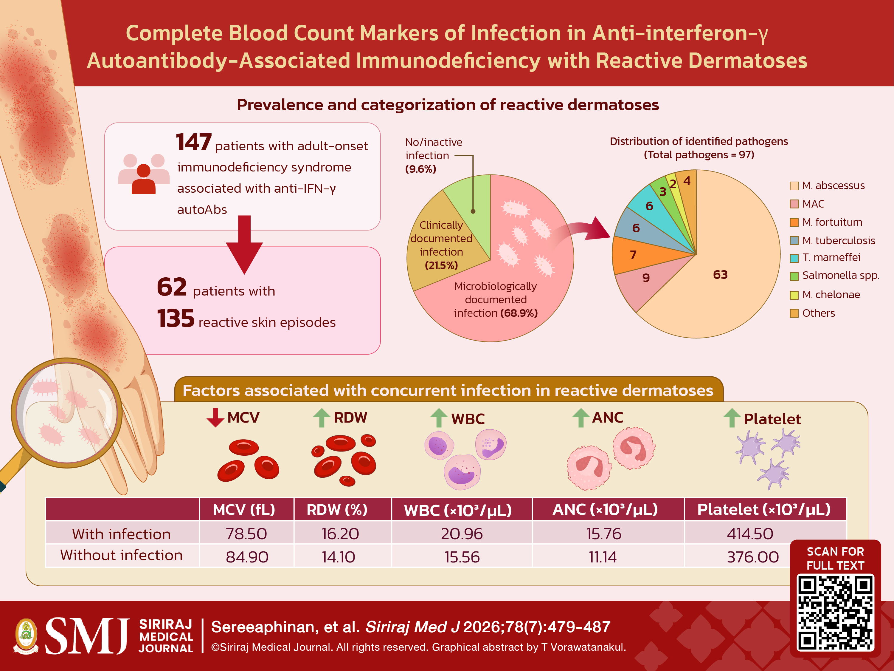
Results: Of the 147 patients reviewed, 62 (42.2%) presented with reactive dermatoses, accounting for 135 episodes. Patients with concurrent infections had significantly lower mean corpuscular volume (78.5 vs. 84.9 fL; p=0.039). In contrast, red cell distribution width (16.2 vs. 14.1%; p=0.005), white blood cell count (20.96 vs. 15.56 ×10³/μL; p=0.037), absolute neutrophil count (15.76 vs. 11.14 ×10³/μL; p=0.038), and platelet count (414.5 vs. 376.0 ×10³/μL; p=0.023), were significantly higher in infected episodes than in non-infected episodes.
Conclusion: In patients presenting with reactive dermatoses, laboratory parameters derived from the routine complete blood count can serve as valuable indicators of increased risk for concurrent infections, enabling prompt diagnosis and targeted management.
References
Yi P, Jiang J, Wang Z, Wang X, Zhao M, Wu H, et al. Comparison of mean platelet volume (MPV) and red blood cell distribution width (RDW) between psoriasis patients and controls: A systematic review and meta-analysis. PLoS One. 2022;17(2):e0264504.
Kim DS, Shin D, Kim TG, Kim SH, Kim DY, Kim SM, et al. Red blood cell distribution width as a useful indicator to predict systemic vasculitis in patients with cutaneous vasculitis. Rheumatol Int. 2015;35(4):719-25.
Şener G, İnan Yuksel E, Gökdeniz O, Karaman K, Canat HD. The Relationship of Hematological Parameters and C-reactive Protein (CRP) With Disease Presence, Severity, and Response to Systemic Therapy in Patients With Psoriasis. Cureus. 2023;15(8):e43790.
Tungphaisal V, Phinyo P, Rujiwetpongstorn R, Kiratikanon S, Tovanabutra N, Chaiwarith R, et al. Reactive Neutrophilic Dermatoses in Adult-Onset Immunodeficiency due to Interferon-Gamma Autoantibody and Their Associated Factors. Dermatology. 2023;239(2):248-54.
Shih HP, Ding JY, Yeh CF, Chi CY, Ku CL. Anti-interferon-γ autoantibody-associated immunodeficiency. Curr Opin Immunol. 2021;72:206-14.
Jutivorakool K, Sittiwattanawong P, Kantikosum K, Hurst CP, Kumtornrut C, Asawanonda P, et al. Skin Manifestations in Patients with Adult-onset Immunodeficiency due to Anti-interferon-gamma Autoantibody: A Relationship with Systemic Infections. Acta Derm Venereol. 2018;98(8):742-7.
Angkasekwinai N, Suputtamongkol Y, Phoompoung P, Pithukpakorn M, Wongswat E, Umrod P, et al. Clinical outcome and laboratory markers for predicting disease activity in patients with disseminated opportunistic infections associated with anti-interferon-γ autoantibodies. PLoS One. 2019;14(4):e0215581.
Pattanaprichakul P, Leeyaphan C, Angkasekwinai N, Bunyaratavej S, Senawong S, Sereeaphinan C, et al. Prevalence and clinical manifestations of cutaneous findings in patients with adult-onset immunodeficiency due to anti-interferon gamma autoantibodies: an eight-year retrospective study. Int J Dermatol. 2023;62(12):1506-10.
Ding L, Mo A, Jutivorakool K, Pancholi M, Holland SM, Browne SK. Determination of human anticytokine autoantibody profiles using a particle-based approach. J Clin Immunol. 2012;32(2):238-45.
Kobkurkul P PC, Rueangkaew J, Angkoolpakdeekul N, Varothai S, Bunyaratavej S, Silpa-archa N. Retrospective Analysis of Inpatient Dermatologic Consultations in a Residency Training Program. Siriraj Med J. 2024;76(2):52-60.
Bunyaratavej S, Jirawattanadon P, Sereeaphinan C, Wongdama S, Sombatmaithai S, Leeyaphan C. Mobile Device Digital Photography for Teledermatology Consultation: Real-Life Situations. Siriraj Med J. 2023;75(12):871–9.
Kiratikanon S, Phinyo P, Rujiwetpongstorn R, Patumanond J, Tungphaisal V, Mahanupab P, et al. Adult-onset immunodeficiency due to anti-interferon-gamma autoantibody-associated Sweet syndrome: A distinctive entity. J Dermatol. 2022;49(1):133-41.
Chi CY, Lin CH, Ho MW, Ding JY, Huang WC, Shih HP, et al. Clinical manifestations, course, and outcome of patients with neutralizing anti-interferon-γ autoantibodies and disseminated nontuberculous mycobacterial infections. Medicine (Baltimore). 2016;95(25):e3927.
Wongkulab P, Wipasa J, Chaiwarith R, Supparatpinyo K. Autoantibody to interferon-gamma associated with adult-onset immunodeficiency in non-HIV individuals in Northern Thailand. PLoS One. 2013;8(9):e76371.
Leeyaphan J, Sutha P, Thawaornwan U, Muennoo T. Vertebral Osteomyelitis Due to Mycobacterium abscessus in an HIV-Negative Patient: Case Report and Literature Review. Siriraj Med J. 2014;2(66):49-52.
Keel SB, Abkowitz JL. The microcytic red cell and the anemia of inflammation. N Engl J Med. 2009;361(19):1904-6.
Liu Q, Dang AM, Chen BW, Lv NQ, Wang X, Zheng DY. The association of red blood cell distribution width with anemia and inflammation in patients with Takayasu arteritis. Clin Chim Acta. 2015;438:205-9.
Hu ZD, Chen Y, Zhang L, Sun Y, Huang YL, Wang QQ, et al. Red blood cell distribution width is a potential index to assess the disease activity of systemic lupus erythematosus. Clin Chim Acta. 2013;425:202-5.
Linden MD. Platelet physiology. Methods Mol Biol. 2013;992:13-30.
McNicol A, Israels SJ. Beyond hemostasis: the role of platelets in inflammation, malignancy and infection. Cardiovasc Hematol Disord Drug Targets. 2008;8(2):99-117.
Liu S, Zhao K, Shao C, Xu L, Cui X, Wang Y. Association between alkaline phosphatase to albumin ratio and mortality among patients with sepsis. Sci Rep. 2024;14(1):3170.
Katasako A, Sasaki S, Raita Y, Yamamoto S, Tochitani K, Murakami M, et al. Association between serum alkaline phosphatase and bacteraemia in haemodialysis outpatients: a multicentre retrospective cross-sectional study. BMJ Open. 2022;12(10):e058666.
Zhang H, Xie S, Li Y, Li J, Deng P, Zeng H, et al. The potential performance of serum albumin to globulin ratio, albumin and globulin in the diagnosis of periprosthetic joint infection and prediction of reinfection following reimplantation. BMC Musculoskelet Disord. 2022;23(1):730.
Published
How to Cite
License
Copyright (c) 2026 Siriraj Medical Journal

This work is licensed under a Creative Commons Attribution-NonCommercial-NoDerivatives 4.0 International License.
Authors who publish with this journal agree to the following conditions:
Copyright Transfer
In submitting a manuscript, the authors acknowledge that the work will become the copyrighted property of Siriraj Medical Journal upon publication.
License
Articles are licensed under a Creative Commons Attribution-NonCommercial-NoDerivatives 4.0 International License (CC BY-NC-ND 4.0). This license allows for the sharing of the work for non-commercial purposes with proper attribution to the authors and the journal. However, it does not permit modifications or the creation of derivative works.
Sharing and Access
Authors are encouraged to share their article on their personal or institutional websites and through other non-commercial platforms. Doing so can increase readership and citations.